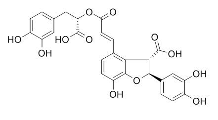
Lithospermic acid
Lithospermic acid has anti-HIV, antioxidant ,anti-inflammatory, and hepatoprotective effects, is a competitive inhibitor of xanthine oxidas (XO), can directly scavenge superoxide and inhibit superoxide production in vitro, and presents hypouricemic actions in vivo. Lithospermic acid has inhibitory effects on proliferation and migration of rat vascular smooth muscle cells, it has a preventive effect on the development of diabetic retinopathy. Lithospermic acid can attenuate 1-methyl-4-phenylpyridine-induced neurotoxicity by blocking neuronal apoptotic and neuroinflammatory pathways. Lithospermic acid can attenuate mesenteric ischemia reperfusion injury in rat intestines by increasing tissue SOD and GPx activities and decreasing MDA and MPO levels, also improves morphological alterations which occurred after periods of reperfusion.
Inquire / Order:
manager@chemfaces.com
Technical Inquiries:
service@chemfaces.com
Tel:
+86-27-84237783
Fax:
+86-27-84254680
Address:
1 Building, No. 83, CheCheng Rd., Wuhan Economic and Technological Development Zone, Wuhan, Hubei 430056, PRC
Providing storage is as stated on the product vial and the vial is kept tightly sealed, the product can be stored for up to
24 months(2-8C).
Wherever possible, you should prepare and use solutions on the same day. However, if you need to make up stock solutions in advance, we recommend that you store the solution as aliquots in tightly sealed vials at -20C. Generally, these will be useable for up to two weeks. Before use, and prior to opening the vial we recommend that you allow your product to equilibrate to room temperature for at least 1 hour.
Need more advice on solubility, usage and handling? Please email to: service@chemfaces.com
The packaging of the product may have turned upside down during transportation, resulting in the natural compounds adhering to the neck or cap of the vial. take the vial out of its packaging and gently shake to let the compounds fall to the bottom of the vial. for liquid products, centrifuge at 200-500 RPM to gather the liquid at the bottom of the vial. try to avoid loss or contamination during handling.
Free Radic Biol Med.2021, 166:104-115.
J Ethnopharmacol.2018, 210:88-94
Int J Mol Sci.2023, 24(18):13713.
Molecules.2021, 26(12):3652.
Evid Based Complement Alternat Med.2015, 2015:165457
Phytomedicine.2021, 2(82):153452
Environ Toxicol.2024, tox.24246
Rep.Grant.Res.,Asahi Glass Foun.2023, No.119.
Jeju National University Graduate School2023, 24478
Buildings2023, 13(5), 1112.
Related and Featured Products
Adv Clin Exp Med. 2012 Jul-Aug;21(4):433-9.
Lithospermic acid and ischemia/reperfusion injury of the rat small intestine prevention.[Pubmed:
23240448]
Intestinal ischemia and reperfusion (I-R) injury of different causes, including cardiac insufficiency, sepsis, vasodepressant and cardiodepressant drugs, and complications of long-lasting surgery, represents a major clinical problem.
The purpose of the present study was to investigate whether Lithospermic acid (LA) can reduce oxidative stress and histological damage in the rat small bowel subjected to mesenteric I-R injury.
METHODS AND RESULTS:
The study was performed on three groups of animals, each composed of 7 rats: the SO (sham operation) group, the I-R/Untreated group and the I-R/LA (I-R plus LA pretreatment) group. Intestinal ischemia for 45 minutes and reperfusion for 60 minutes were applied. Ileum specimens were obtained to determine the tissue level of malondialdehyde (MDA), superoxide dismutase (SOD), glutathione peroxidase (GPx), catalase (CAT) and myeloperoxidase (MPO) activities and histological changes.
Untreated intestinal I-R resulted in increased tissue MDA and MPO levels and diminished SOD and GPx activities. These changes were found to be almost reversed in the LA treatment group. Histopathologically, the intestinal injury in rats treated with LA was less than the untreated I-R group.
CONCLUSIONS:
Lithospermic acid attenuates mesenteric ischemia reperfusion injury in rat intestines by increasing tissue SOD and GPx activities and decreasing MDA and MPO levels. Lithospermic acid also improves morphological alterations which occurred after periods of reperfusion.
PLoS One. 2014 Jun 6;9(6):e98232.
The effect of lithospermic acid, an antioxidant, on development of diabetic retinopathy in spontaneously obese diabetic rats.[Pubmed:
24905410]
Lithospermic acid B (LAB), an active component isolated from Salvia miltiorrhiza radix, has been reported to have antioxidant effects. We examined the effects of LAB on the prevention of diabetic retinopathy in Otsuka Long-Evans Tokushima Fatty (OLETF) rats, an animal model of type 2 diabetes.
METHODS AND RESULTS:
LAB (10 or 20 mg/kg) or normal saline were given orally once daily to 24-week-old male OLETF rats for 52 weeks. At the end of treatment, fundoscopic findings, vascular endothelial growth factor (VEGF) expression in the eyeball, VEGF levels in the ocular fluid, and any structural abnormalities in the retina were assessed. Glucose metabolism, serum levels of high-sensitivity C-reactive protein (hsCRP), monocyte chemotactic protein-1 (MCP1), and tumor necrosis factor-alpha (TNFα) and urinary 8-hydroxy-2'-deoxyguanosine (8-OHdG) levels were also measured. Treatment with LAB prevented vascular leakage and basement membrane thickening in retinal capillaries in a dose-dependent manner. Insulin resistance and glucose intolerance were significantly improved by LAB treatment. The levels of serum hsCRP, MCP1, TNFα, and urinary 8-OHdG were lower in the LAB-treated OLETF rats than in the controls.
CONCLUSIONS:
Treatment with LAB had a preventive effect on the development of diabetic retinopathy in this animal model, probably because of its antioxidative effects and anti-inflammatory effects.
Chem Biol Interact. 2008 Nov 25;176(2-3):137-42.
Lithospermic acid as a novel xanthine oxidase inhibitor has anti-inflammatory and hypouricemic effects in rats.[Pubmed:
18694741 ]
Lithospermic acid (LSA) was originally isolated from the roots of Salvia mitiorrhiza, a common herb of oriental medicine. Previous studies demonstrated that LSA has antioxidant effects. In this study, we investigated the in vitro xanthine oxidase (XO) inhibitory activity, and in vivo hypouricemic and anti-inflammatory effects of rats.
METHODS AND RESULTS:
XO activity was detected by measuring the formation of uric acid or superoxide radicals in the xanthine/xanthine oxidase system. The results showed that LSA inhibited the formation of uric acid and superoxide radicals significantly with an IC50 5.2 and 1.08 microg/ml, respectively, and exhibited competitive inhibition. It was also found that LSA scavenged superoxide radicals directly in the system beta-NADH/PMS and inhibited the production of superoxide in human neutrophils stimulated by PMA and fMLP. LSA was also found to have hypouricemic activity on oxonate-pretreated rats in vivo and have anti-inflammatory effects in a model of gouty arthritis.
CONCLUSIONS:
These results suggested that LSA is a competitive inhibitor of XO, able to directly scavenge superoxide and inhibit superoxide production in vitro, and presents hypouricemic and anti-inflammatory actions in vivo.
Oncol Rep . 2015 Aug;34(2):673-80.
Anti-oxidative and hepatoprotective effects of lithospermic acid against carbon tetrachloride-induced liver oxidative damage in vitro and in vivo[Pubmed:
26081670]
Abstract
Accumulation of an excess amount of reactive oxygen species (ROS) can cause hepatotoxicity that may result in liver damage. Therefore, development of anti-oxidative agents is needed for reducing liver toxicity. This study investigated the anti-oxidative and hepatoprotective activity of Lithospermic acid, a plant-derived polycyclic phenolic carboxylic acid isolated from Salvia miltiorrhiza, on carbon tetrachloride (CCl4)-induced acute liver damage in vitro and in vivo. The results of the DPPH assay indicated that Lithospermic acid was a good anti-oxidant. the CCl4-exposed Huh7 cell line exhibited decreased cell viability, increased necrosis and elevated ROS and caspase-3/7 activity. Lithospermic acid significantly attenuated the CCl4-induced oxidative damage in a concentration-dependent manner. The result of an in vivo study with BALB/c mice corresponded with the anti-oxidative activity noted in the in vitro study. Exposure of mice to CCl4 resulted in a greater than 2-fold elevation in serum aspartate transaminase (AST) and alanine transaminase (ALT). levels In addition, CCl4-intoxication led to an over 20% decrease in the level of intracellular hepatic enzymes including superoxide dismutase (SOD) and catalase (CAT) as well as increased lipid peroxidation. Upon histological examination of the CCl4-exposed mice, the mouse livers showed severe hepatic damage with a huge section of necrosis and structural destruction. Pretreatment of mice with Lithospermic acid for six days significantly reduced CCl4-induced hepatic oxidative damage, serum AST and ALT. The pretreatment also increased SOD and CAT. The findings suggest that the health status of the liver was improved comparable to the control group after a high-dose treatment with Lithospermic acid (100 mg/kg weight). The potential applicability of Lithospermic acid as a hepatoprotective agent was demonstrated.
Acta Pharmacol Sin. 2009 Sep;30(9):1245-52.
Inhibitory effects of lithospermic acid on proliferation and migration of rat vascular smooth muscle cells.[Pubmed:
19701233]
To understand the effects of Lithospermic acid (LA), a potent antioxidant from the water-soluble extract of Salvia miltiorrhiza, on the migration and proliferation of rat thoracic aorta vascular smooth muscle cells (VSMCs).
METHODS AND RESULTS:
VSMC migration, proliferation, DNA synthesis and cell cycle progression were investigated by transwell migration analysis, 3-(4,5-dimethylthiazol-2-yl)-2,5-diphenyltetrazolium bromide (MTT) assay, bromodeoxyuridine (BrdU) incorporation assay, and flow cytometric detection, respectively. Intracellular reactive oxygen species (ROS) generation was detected using 2',7'-dichlorofluorescin diacetate (DCFH-DA). The expression of cyclin D1 protein and matrix metalloproteinase-9 (MMP-9) protein, as well as the phosphorylation state of ERK1/2, were determined using Western blots. The activity of MMP-9 and the expression of MMP-9 mRNA were assessed by gelatin zymography analysis and RT-PCR, respectively.
LA (25-100 micromol/L) inhibited both lipopolysaccharide (LPS)- and fetal bovine serum (FBS)-induced ROS generation and ERK1/2 phosphorylation. By down-regulating the expression of cyclin D(1) and arresting cell cycle progression at the G(1) phase, LA inhibited both VSMC proliferation and DNA synthesis as induced by 5% FBS. Furthermore, LA attenuated LPS-induced VSMC migration by inhibiting MMP-9 expression and its enzymatic activity.
CONCLUSIONS:
LA is able to inhibit FBS-induced VSMC proliferation and LPS-induced VSMC migration, which suggests that LA may have therapeutic effects in the prevention of atherosclerosis, restenosis and neointimal hyperplasia.
J Biomed Sci. 2015 May 28;22(1):37.
Lithospermic acid attenuates 1-methyl-4-phenylpyridine-induced neurotoxicity by blocking neuronal apoptotic and neuroinflammatory pathways.[Pubmed:
26018660]
Parkinson's disease is the second most common neurodegenerative disorders after Alzheimer's disease. The main cause of the disease is the massive degeneration of dopaminergic neurons in the substantia nigra. Neuronal apoptosis and neuroinflammation are thought to be the key contributors to the neuronal degeneration.
METHODS AND RESULTS:
Both CATH.a cells and ICR mice were treated with 1-methyl-4-phenylpyridin (MPP(+)) to induce neurotoxicity in vitro and in vivo. Western blotting and immunohistochemistry were also used to analyse neurotoxicity, neuroinflammation and aberrant neurogenesis in vivo. The experiment in CATH.a cells showed that the treatment of MPP(+) impaired intake of cell membrane and activated caspase system, suggesting that the neurotoxic mechanisms of MPP(+) might include both necrosis and apoptosis. Pretreatment of Lithospermic acid might prevent these toxicities. Lithospermic acid possesses specific inhibitory effect on caspase 3. In mitochondria, MPP(+) caused mitochondrial depolarization and induced endoplasmic reticulum stress via increasing expression of chaperone protein, GRP-78. All the effects mentioned above were reduced by Lithospermic acid. In animal model, the immunohistochemistry of mice brain sections revealed that MPP(+) decreased the amount of dopaminergic neurons, enhanced microglia activation, promoted astrogliosis in both substantia nigra and hippocampus, and MPP(+) provoked the aberrant neurogenesis in hippocampus. Lithospermic acid significantly attenuates all of these effects induced by MPP(+).
CONCLUSIONS:
Lithospermic acid is a potential candidate drug for the novel therapeutic intervention on Parkinson's disease.
Oncol Rep. 2015 Aug;34(2):673-80.
Anti-oxidative and hepatoprotective effects of lithospermic acid against carbon tetrachloride-induced liver oxidative damage in vitro and in vivo.[Pubmed:
26081670 ]
Accumulation of an excess amount of reactive oxygen species (ROS) can cause hepatotoxicity that may result in liver damage. Therefore, development of anti-oxidative agents is needed for reducing liver toxicity.
METHODS AND RESULTS:
This study investigated the anti-oxidative and hepatoprotective activity of Lithospermic acid, a plant-derived polycyclic phenolic carboxylic acid isolated from Salvia miltiorrhiza, on carbon tetrachloride (CCl4)-induced acute liver damage in vitro and in vivo. The results of the DPPH assay indicated that Lithospermic acid was a good anti-oxidant. the CCl4-exposed Huh7 cell line exhibited decreased cell viability, increased necrosis and elevated ROS and caspase-3/7 activity. Lithospermic acid significantly attenuated the CCl4-induced oxidative damage in a concentration-dependent manner. The result of an in vivo study with BALB/c mice corresponded with the anti-oxidative activity noted in the in vitro study. Exposure of mice to CCl4 resulted in a greater than 2-fold elevation in serum aspartate transaminase (AST) and alanine transaminase (ALT). levels In addition, CCl4-intoxication led to an over 20% decrease in the level of intracellular hepatic enzymes including superoxide dismutase (SOD) and catalase (CAT) as well as increased lipid peroxidation. Upon histological examination of the CCl4-exposed mice, the mouse livers showed severe hepatic damage with a huge section of necrosis and structural destruction. Pretreatment of mice with Lithospermic acid for six days significantly reduced CCl4-induced hepatic oxidative damage, serum AST and ALT. The pretreatment also increased SOD and CAT.
CONCLUSIONS:
The findings suggest that the health status of the liver was improved comparable to the control group after a high-dose treatment with Lithospermic acid (100 mg/kg weight). The potential applicability of Lithospermic acid as a hepatoprotective agent was demonstrated.
Org Biomol Chem. 2012 Jul 28;10(28):5456-65.
Synthesis of anti-HIV lithospermic acid by two diverse strategies.[Pubmed:
22669348]
An efficient and convergent route for the synthesis of the natural product (+)-Lithospermic acid, which possesses anti-HIV activity, was accomplished. The (±)-trans-dihydrobenzo[b]furan core therein was prepared by two different strategies.
METHODS AND RESULTS:
The first strategy involved the use of a palladium-catalyzed annulation to generate an appropriately substituted benzo[b]furan ester followed by a stereoselective reduction of a carbon-carbon double bond with Mg-HgCl(2)-MeOH. The second strategy relied on an aldol condensation between a suitably substituted methyl arylacetate and 3,4-dimethoxybenzaldehyde, followed by cyclization.
CONCLUSIONS:
Finally, a total synthesis of (+)-Lithospermic acid was completed via coupling of a trans-dihydrobenzo[b]furan cinnamic acid with an enantiomerically pure methyl lactate.
Bioorg. Med. Chem. Lett., 2009, 19(6):1815-7.
Lithospermic acid derivatives from Lithospermum erythrorhizon increased expression of serine palmitoyltransferase in human HaCaT cells.[Pubmed:
19217780 ]
A MeOH extract of the dry root of Lithospermum erythrorhizon showed strong increasing effect on serine palmitoyltransferase (SPT) in normal human keratinocyte cells (HaCaT cells).
METHODS AND RESULTS:
Bioassay-guided separation on this extract using repeated chromatography resulted in the isolation of Lithospermic acid (1) and two derivative esters, 9''-methyl lithospermate (2) and 9'-methyl lithospermate (3). Compounds 1-3 significantly increased SPT expressions in the relative quantity (%) of SPT1 mRNA as well as SPT2 mRNA. These constituents also raised the level of SPT protein in HaCaT cells in a dose-dependent manner, with the increased level of SPT protein in HaCaT cells of 55%, 23%, and 81% at the concentration of 100 microg/ml, respectively.
CONCLUSIONS:
This finding suggests that Lithospermic acid and its derivatives from L. erythrorhizon might improve the permeability barrier by stimulating the protein level of SPT.


