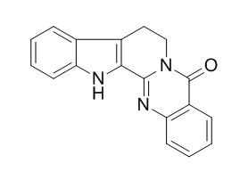
Rutaecarpine
Rutaecarpine is an inhibitor of COX-2 with an IC50 value of 0.28 μM, and is also a potent inhibitor of CYP1A2. Rutaecarpine has anti-atherosclerosis, immunosuppressive, anti-inflammatory, gastroprotective, vasorelaxing, antihypertensive and anti-platelet effects. Rutaecarpine has positive inotropic and chronotropic effects on the guinea-pig isolated right atria, possible involvement of vanilloid receptors. Rutaecarpine may be useful in the prevention of ultraviolet A-induced photoaging, it inhibits ultraviolet A-induced reactive oxygen species generation, resulting in the enhanced expression of matrix metalloproteinase (MMP)-2 and MMP-9 in human skin cells.
Inquire / Order:
manager@chemfaces.com
Technical Inquiries:
service@chemfaces.com
Tel:
+86-27-84237783
Fax:
+86-27-84254680
Address:
1 Building, No. 83, CheCheng Rd., Wuhan Economic and Technological Development Zone, Wuhan, Hubei 430056, PRC
Providing storage is as stated on the product vial and the vial is kept tightly sealed, the product can be stored for up to
24 months(2-8C).
Wherever possible, you should prepare and use solutions on the same day. However, if you need to make up stock solutions in advance, we recommend that you store the solution as aliquots in tightly sealed vials at -20C. Generally, these will be useable for up to two weeks. Before use, and prior to opening the vial we recommend that you allow your product to equilibrate to room temperature for at least 1 hour.
Need more advice on solubility, usage and handling? Please email to: service@chemfaces.com
The packaging of the product may have turned upside down during transportation, resulting in the natural compounds adhering to the neck or cap of the vial. take the vial out of its packaging and gently shake to let the compounds fall to the bottom of the vial. for liquid products, centrifuge at 200-500 RPM to gather the liquid at the bottom of the vial. try to avoid loss or contamination during handling.
J Enzyme Inhib Med Chem.2019, 34(1):134-143
Academic J of Second Military Medical University2018, 39(11)
Hum Exp Toxicol.2023, 42:9603271221145386.
Horticulture Research2022, uhac276.
Journal of Molecular Liquids2022, 364:120062.
Neuropharmacology.2018, 131:68-82
Environ Toxicol.2023, 38(7):1641-1650.
Front Pharmacol.2020, 11:683.
J Nat Prod.2023, 86(2):264-275.
Korean J. of Food Sci. and Tech2016, 172-177
Related and Featured Products
Chin J Integr Med. 2014 Sep;20(9):682-7.
Rutaecarpine inhibits angiotensin II-induced proliferation in rat vascular smooth muscle cells.[Pubmed:
23775171]
OBJECTIVE:
To evaluate the effects and possible mechanisms of Rutaecarpine on angiotensin II (Ang II)-induced proliferation in cultured rat vascular smooth muscle cells (VSMCs).
METHODS:
To examine the mechanisms involved in anti-proliferative effects of Rutaecarpine, nitric oxide (NO) levels and NO synthetase (NOS) activity were determined. Expressions of VSMC proliferation-related genes including endothelial nitric oxide synthase (eNOS), and c-myc hypertension related gene-1 (HRG-1) were determined by real-time reverse transcription-polymerase chain reaction (RT-PCR).
RESULTS:
Rutaecarpine (0.3-3.0 μmol/L) inhibited Ang II-induced VSMC proliferation and the best effects were achieved at 3.0 μmol/L. The Ang II-induced decreases in cellular NO contents and NOS activities were antagonized by Rutaecarpine (P <0.05). Ang II administration suppressed the expressions of eNOS and HRG-1, while increased c-myc expression (P <0.05). All these effects were attenuated by 3.0 μmol/L Rutaecarpine (P <0.05).
CONCLUSION:
Rutaecarpine is effective against Ang II-induced rat VSMC proliferation, and this effect is due, at least in part, to NO production and the modulation of VMSC proliferation-related gene expressions.
Drug Metab Dispos. 2002 Mar;30(3):349-53.
The alkaloid rutaecarpine is a selective inhibitor of cytochrome P450 1A in mouse and human liver microsomes.[Pubmed:
11854157]
Rutaecarpine, evodiamine, and dehydroevodiamine are quinazolinocarboline alkaloids isolated from a traditional Chinese medicine, Evodia rutaecarpa. The in vitro effects of these alkaloids on cytochrome P450 (P450)-catalyzed oxidations were studied using mouse and human liver microsomes.
METHODS AND RESULTS:
Among these alkaloids, Rutaecarpine showed the most potent and selective inhibitory effect on CYP1A-catalyzed 7-methoxyresorufin O-demethylation (MROD) and 7-ethoxyresorufin O-deethylation (EROD) activities in untreated mouse liver microsomes. The IC(50) ratio of EROD to MROD was 6. For MROD activity, Rutaecarpine was a noncompetitive inhibitor with a K(i) value of 39 +/- 2 nM. In contrast, Rutaecarpine had no effects on benzo[a]pyrene hydroxylation (AHH), aniline hydroxylation, and nifedipine oxidation (NFO) activities. In human liver microsomes, 1 microM Rutaecarpine caused 98, 91, and 77% decreases of EROD, MROD, and phenacetin O-deethylation activities, respectively. In contrast, less than 15% inhibition of AHH, tolbutamide hydroxylation, chlorzoxazone hydroxylation, and NFO activities were observed in the presence of 1 microM Rutaecarpine. To understand the selectivity of inhibition of CYP1A1 and CYP1A2, inhibitory effects of Rutaecarpine were studied using liver microsomes of 3-methylcholanthrene (3-MC)-treated mice and Escherichia coli membrane expressing bicistronic human CYP1A1 and CYP1A2. Similar to the CYP1A2 inhibitor furafylline, Rutaecarpine preferentially inhibited MROD more than EROD and had no effect on AHH in 3-MC-treated mouse liver microsomes. For bicistronic human P450s, the IC(50) value of Rutaecarpine for EROD activity of CYP1A1 was 15 times higher than the value of CYP1A2.
CONCLUSIONS:
These results indicated that Rutaecarpine was a potent inhibitor of CYP1A2 in both mouse and human liver microsomes.
J Pharmacol Exp Ther. 1996 Mar;276(3):1016-21.
The vasorelaxing action of rutaecarpine: direct paradoxical effects on intracellular calcium concentration of vascular smooth muscle and endothelial cells.[Pubmed:
8786530]
We have examined both the hypotensive effect and the mechanism of intracellular Ca++ regulation, underlying Rutaecarpine (Rut)-induced vasodilatation. An i.v. bolus injection of Rut in anesthetized Sprague-Dawley rats produced a dose-dependent hypotensive effect.
METHODS AND RESULTS:
In isolated rat aorta rings, Rut (0.1-3 mu M) inhibited the phasic and tonic responses of norepinephrine- and phyenylephrine-induced contractions, respectively, mainly through an endothelium-dependent mechanism. However, the vasorelaxing effect of Rut (3 microM) persisted in denuded aorta, although to a much less extent than in intact tissue. As determined by the fura-2/AM (1-[2-(5-carboxyoxazol-2-yl)-6-aminobenzofuran-5-oxy]-2-(2'- amino-5'-methylphenoxy)-ethane-N,N,N,N-tetraacetic acid pentaacetoxymethyl ester) method, Rut (10 microM), in the presence of extracellular Ca++, suppressed the KCI-induced increment in the intracellular Ca++ concentration ([Ca++]i) of cultured vascular smooth muscle cells (VSMC). Rut (10 microM) also attenuated the norepinephrine-induced peak rise of [Ca++]i in VSMC placed in Ca++-free solution. On the other hand, Rut (1 and 10 microM) increased the level of [Ca++]i of cultured endothelial cells (EC) in the presence of extracellular Ca++.
CONCLUSIONS:
In conclusion, Rut acts on both VSMC and EC directly. In VSMC, it reduces [Ca++]i through the inhibition of Ca++ influx and Ca++ release from intracellular stores. In EC, Rut augments EC [Ca++]i by increasing Ca++ influx, possibly leading to nitric oxide release. The paradoxical regulation of Ca++ in both VSMC and EC acts simultaneously to cause vasorelaxation which could account, at least in part, for the hypotensive action. This is a most significant and a unique feature of this study.
Peptides. 2008 Oct;29(10):1781-8.
Calcitonin gene-related peptide-mediated antihypertensive and anti-platelet effects by rutaecarpine in spontaneously hypertensive rats.[Pubmed:
18625276 ]
We have previously reported that Chinese traditional medicine Rutaecarpine (Rut) produced a sustained hypotensive effect in phenol-induced and two-kidney, one-clip hypertensive rats. The aims of this study are to determine whether Rut could exert antihypertensive and anti-platelet effects in spontaneously hypertensive rats (SHR) and the underlying mechanisms.
METHODS AND RESULTS:
In vivo, SHR were given Rut and the blood pressure was monitored. Blood was collected for the measurements of calcitonin gene-related peptide (CGRP), tissue factor (TF) concentration and activity, and platelet aggregation, and the dorsal root ganglia were saved for examining CGRP expression. In vitro, the effects of Rut and CGRP on platelet aggregation were measured, and the effect of CGRP on platelet-derived TF release was also determined. Rut exerted a sustained hypotensive effect in SHR concomitantly with the increased synthesis and release of CGRP. The treatment of Rut also showed an inhibitory effect on platelet aggregation concomitantly with the decreased TF activity and TF antigen level in plasma. Study in vitro showed an inhibitory effect of Rut on platelet aggregation in the presence of thoracic aorta, which was abolished by capsazepine or CGRP(8-37), an antagonist of vanilloid receptor or CGRP receptor. Exogenous CGRP was able to inhibit both platelet aggregation and the release of platelet-derived TF, which were abolished by CGRP(8-37).
CONCLUSIONS:
The results suggest that Rut exerts both antihypertensive and anti-platelet effects through stimulating the synthesis and release of CGRP in SHR, and CGRP-mediated anti-platelet effect is related to inhibiting the release of platelet-derived TF.
Toxicol Lett. 2006 Jul 1;164(2):155-66.
Immunosuppressive effects of rutaecarpine in female BALB/c mice.[Pubmed:
16412592]
Rutaecarpine is a major quinazolinocarboline alkaloid isolated from Evodia rutaecarpa. It was reported to possess a wide spectrum of pharmacological activities, such as vasodilation, antithrombosis, and anti-inflammation.
METHODS AND RESULTS:
In the present study, adverse effects of Rutaecarpine on immune functions were determined in female BALB/c mice. Rutaecarpine had no effects on hepatotoxicity parameters in mice, as measured by serum activities of aminotransferases. Meanwhile, Rutaecarpine significantly decreased the number of antibody-forming cells and caused weight decrease in spleen in a dose-dependent manner, when mice were administered with Rutaecarpine at 10mg/kg, 20mg/kg, 40 mg/kg or 80 cmg/kg once intravenously. In addition, Rutaecarpine administered mice exhibited reduced splenic cellularity, decreased numbers of total T cells, CD4(+) cells, CD8(+) cells, and B cells in spleen. IL-2, interferon-gamma and IL-10 mRNA expressions were suppressed significantly by Rutaecarpine treatment. The number of CD4(+)IL-2(+) cells was reduced significantly following administration of mice with Rutaecarpine. Furthermore, Rutaecarpine caused the cell cycle arrest in G(0)+G(1) phase in a dose-dependent manner. Rutaecarpine caused significant inductions of hepatic cytochrome P450 (CYP) 1A, 2B, and 2E1 activities dose-dependently. In the splenic lymphocyte proliferation assay, Rutaecarpine inhibited proliferation by LPS and Con A ex vivo, whereas it had no effects on in vitro proliferation.
CONCLUSIONS:
These results suggested that a single bolus intravenous injection of Rutaecarpine from 20mg/kg might cause immunosuppressive effects, and that Rutaecarpine-induced immunosuppression might be mediated, at least in part, through the inhibition of cytokine production and cell cycle arrest in G(0)+G(1) phase, and caused possibly by mechanisms associated with metabolic activation.
Inflamm Res. 1999 Dec;48(12):621-5.
A new class of COX-2 inhibitor, rutaecarpine from Evodia rutaecarpa.[Pubmed:
10669112 ]
We investigated the effect of a new class of COX-2 inhibitor, Rutaecarpine, on the production of PGD2 in bone marrow derived mast cells (BMMC) and PGE2 in COX-2 transfected HEK293 cells. Inflammation was induced by lambda-carrageenan in male Splague-Dawley (SD) rats.
METHODS AND RESULTS:
Rutaecarpine (8,13-Dihydroindolo[2',3':3,4]pyridol[2,1-b]quinazolin -5(7H)-one) was isolated from the fruits of Evodia rutaecarpa. BMMC were cultured with WEHI-3 conditioned medium. c-Kit ligand and IL-10 were obtained by their expression in baculovirus.
The generation of PGD2 and PGE2 were determined by their assay kit. COX-1 and COX-2 protein and mRNA expression was determined by BMMC in the presence of KL, LPS and IL-10.
Rutaecarpine and indomethacin dissolved in 0.1% carboxymethyl cellulose was administered intraperitoneally and, 1 h later, lambda-carrageenan solution was injected to right hind paw of rats. Paw volumes were measured using plethysmometer 5 h after lambda-carrageenan injection.
Rutaecarpine inhibited COX-2 and COX-1 dependent phases of PGD2 generation in BMMC in a concentration-dependent manner with an IC50 of 0.28 microM and 8.7 microM, respectively. It inhibited COX-2-dependent conversion of exogenous arachidonic acid to PGE2 in a dose-dependent manner by the COX-2-transfected HEK293 cells. However, Rutaecarpine inhibited neither PLA2 and COX-1 activity nor COX-2 protein and mRNA expression up to the concentration of 30 microM in BMMC, indicating that Rutaecarpine directly inhibited COX-2 activity. Furthermore, Rutaecarpine showed in vivo anti-inflammatory activity on rat lambda-carrageenan induced paw edema by intraperitoneal administration.
CONCLUSIONS:
Anti-inflammatory activity of Evodia rutaecarpa could be attributed at least in part by inhibition of COS-2.
Eur J Pharmacol. 2015 Jun 5;756:8-14.
Rutaecarpine prevented dysfunction of endothelial gap junction induced by Ox-LDL via activation of TRPV1.[Pubmed:
25794845]
Gap junctions, which is formed by connexins, has been proved to play an important role in the atherogenesis development. Rutaecarpine was reported to inhibited monocyte migration, which indicates its potential for anti-atherosclerosis activity.
This study evaluated the effect of Rutaecarpine on endothelial dysfunction, and focused on the regulation of connexin expression in endothelial cells by Rutaecarpine.
METHODS AND RESULTS:
Endothelia damage was induced by exposing HUVEC-12 to Ox-LDL (100mg/l) for 24h, which decreased the expression of protective proteins Cx37 and Cx40, but induced atherogenic Cx43 expression, in both mRNA and protein levels, concomitant with the impaired propidium iodide diffusion through the gap junctions. Pretreatment with Rutaecarpine effectively recovered the expression of Cx37 and Cx40, but inhibited Cx43 expression, thereby improving gap junction communication and significantly prevented the endothelial dysfunction. Consequently, the cell viability and nitric oxide production were increased, lactate dehydrogenase production was decreased and monocyte adhesion was inhibited. These protective effects of Rutaecarpine were remarkably attenuated by pretreatment with capsazepine, a competitive antagonist of transient receptor potential vanilloid subtype 1 (TRPV1).
CONCLUSIONS:
In summary, this study is the first to report that Rutaecarpine prevents endothelial injury and gap junction dysfunction induced by Ox-LDL in vitro, which is related to regulation of connexin expression patterns via TRPV1 activation. These results suggest that Rutaecarpine has the potential for use as an anti-atherosclerosis agent with a novel mechanism.
Eur J Pharmacol. 2004 Sep 13;498(1-3):19-25.
Inhibition of UVA irradiation-modulated signaling pathways by rutaecarpine, a quinazolinocarboline alkaloid, in human keratinocytes.[Pubmed:
15363971 ]
Matrix metalloproteinases (MMPs), a key component in photoaging of the skin due to exposure to ultraviolet A, appear to be increased by ultraviolet A irradiation-associated generation of reactive oxygen species.
METHODS AND RESULTS:
In this study, we investigated the effects of synthetic Rutaecarpine, which is also found in Evodia rutaecarpa, on the ultraviolet A-induced changes in the expression of gelatinases: matrix metalloproteinase (MMP)-2 and MMP-9 using HaCaT human keratinocytes as a model cellular system. Ultraviolet A irradiation of HaCaT cells increased the gelatinolytic activities of MMP-2 and MMP-9, which was significantly suppressed by the pretreatment with Rutaecarpine. In addition, Rutaecarpine significantly suppressed the ultraviolet A-induced enhanced expression of MMP-2 and MMP-9 proteins and mRNAs. Rutaecarpine also inhibited the H2O2-induced increase in the expression of MMP-2 and MMP-9. Furthermore, Rutaecarpine decreased the ultraviolet A-induced increased generation of reactive oxygen species.
CONCLUSIONS:
Taken together, these results suggest that Rutaecarpine inhibited ultraviolet A-induced reactive oxygen species generation, resulting in the enhanced expression of MMP-2 and MMP-9 in human skin cells.
These results further suggest that ruetaecarpine may be useful in the prevention of ultraviolet A-induced photoaging.
Zhongguo Zhong Yao Za Zhi. 2014 Aug;39(15):2930-5.
Effects of rutaecarpine on inflammatory cytokines in insulin resistant primary skeletal muscle cells.[Pubmed:
25423835]
It is now well established that inflammation plays an important role in the development of numerous chronic metabolic diseases including insulin resistance (IR) and type 2 diabetes (T2DM). Skeletal muscle is responsible for 75% of total insulin-dependent glucose uptake; consequently, skeletal muscle IR is considered to be the primary defect of systemic IR development.
Our pre- vious study has shown that Rutaecarpine (Rut) can benefit blood lipid profile, mitigate inflammation, and improve kidney, liver, pan- creas pathology status of T2DM rats. However, the effects of Rut on inflammatory cytokines in the development of IR-skeletal muscle cells have not been studied. Thus, our objective was to investigate effects of Rut on inflammatory cytokines interleukiri (IL)-1, IL-6 and tumor necrosis factor (TNF)-α in insulin resistant primary skeletal muscle cells (IR-PSMC).
METHODS AND RESULTS:
Primary cultures of skeletal muscle cells were prepared from 5 neonate SD rats, and the primary rat skeletal muscle cells were identified by cell morphology, effect of ru- taecarpine on cell proliferation by MTT assay. IR-PSMC cells were induced by palmitic acid (PA), the glucose concentration was measured by glucose oxidase and peroxidase (GOD-POD) method. The effects of Rut on inflammatory cytokines IL-1, IL-6 and TNF-α in IR-PSMC cells were tested by enzyme-linked immunosorbent assay (ELISA) kit. The results show that the primary skeletal muscle cells from neonatal rat cultured for 2-4 days, parallel alignment regularly, and cultured for 7 days, cells fused and myotube formed. It was shown that Rut in concentration 0-180. 0 μmol x L(-1) possessed no cytotoxic effect towards cultured primary skeletal muscle cells. However, after 24 h exposure to 0.6 mmol x L(-1) PA, primary skeletal muscle cells were able to induce a state of insulin resistance. The results obtained indicated significant decrease (P < 0.05 to P < 0.001) IL-1, IL-6 and TNF-α production by cultured IR-PSMC cells when incubating 24 hours with Rut, beginning from 20 to 180.0 μmol x L(-1). IL-1, IL-6 and TNF-α in the Rut treated groups were dose-dependently decreased compared with that in the IR-PSMC control group.
CONCLUSIONS:
Our results demonstrated that the Rut promoted glucose consumption and improved insulin resistance possibly through suppression of inflammatory cytokines in the IR-PSMC cells.
Planta Med. 2005 May;71(5):416-9.
The protective effects of rutaecarpine on gastric mucosa injury in rats.[Pubmed:
15931578 ]
Previous investigations have shown that calcitonin gene-related peptide (CGRP) protects gastric mucosa against injury induced by acetylsalicylic acid (ASA) and that Rutaecarpine activates vanilloid receptors to evoke CGRP release.
METHODS AND RESULTS:
In the present study, we examined the protective effects of Rutaecarpine on gastric mucosa injury, and explored whether the protective effects of Rutaecarpine are related to stimulation of endogenous CGRP release via activating vanilloid receptors in rats. In an ASA-induced ulceration model, gastric mucosal ulcer index, pH value of gastric juice and plasma concentrations of CGRP were determined. ASA significantly increased the gastric mucosal ulcer index and the back-diffusion of H+ through the mucosa. Rutaecarpine at the doses of 100 or 300 microg/kg (i.v.), and 300 or 600 microg/kg (intragastric, i.g.) reduced the ulcer index and back-diffusion of H+, which was abolished by pretreatment with capsaicin (50 mg/kg, s.c.) or capsazepine (3 mg/kg, i.v.), a competitive vanilloid receptor antagonist. Rutaecarpine significantly increased the plasma concentration of CGRP, which was also abolished by capsazepine. In a stress-induced ulceration model, Rutaecarpine reduced gastric mucosal damages, which was abolished by capsazepine (5 mg/kg, i.p.).
CONCLUSIONS:
These results suggest that Rutaecarpine protects the gastric mucosa against injury induced by ASA and stress, and that the gastroprotective effect of Rutaecarpine is related to a stimulation of endogenous CGRP release via activation of the vanilloid receptor.
Other References Information
Planta Med. 2001 Apr;67(3):244-8.
The positive inotropic and chronotropic effects of evodiamine and rutaecarpine, indoloquinazoline alkaloids isolated from the fruits of Evodia rutaecarpa, on the guinea-pig isolated right atria: possible involvement of vanilloid receptors.[Pubmed:
11345696]
The effects of evodiamine (1 microM), Rutaecarpine (3 microM) and capsaicin (0.3 microM) were also significantly reduced by pretreatment with ruthenium red (10 microM) and CGRP (8-37) (10 microM). The effects of evodiamine, Rutaecarpine and capsaicin were not affected by pretreatment with PPADS (100 microM), a highly selective P2X purinoceptor antagonist, and the possibility of the involvement of the P2X purinoceptor was excluded. These results suggest that the positive inotropic and chronotropic effects on the guinea-pig isolated right atria induced by both evodiamine and Rutaecarpine could be attributed to their interaction with vanilloid receptors and the resultant release of CGRP, a cardiotonic neurotransmitter, from capsaicin-sensitive nerves as with capsaicin.


