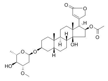
Oleandrin
Oleandrin has anticarcinogenic, anti-inflammatory, and growth-modulatory effects , which may thus be partially ascribed to the inhibition of activation of NF-κB and AP-1 and potentiation of apoptosis; it has stronger anti-proliferative activity in undifferentiated CaCO-2 cells (IC50, 8.25 nM) , causes an autophagic cell death and altered ERK phosphorylation in undifferentiated.Oleandrin inhibits the Na+, K+-ATPase activity with an IC50 of 620 nM.
Inquire / Order:
manager@chemfaces.com
Technical Inquiries:
service@chemfaces.com
Tel:
+86-27-84237783
Fax:
+86-27-84254680
Address:
1 Building, No. 83, CheCheng Rd., Wuhan Economic and Technological Development Zone, Wuhan, Hubei 430056, PRC
Providing storage is as stated on the product vial and the vial is kept tightly sealed, the product can be stored for up to
24 months(2-8C).
Wherever possible, you should prepare and use solutions on the same day. However, if you need to make up stock solutions in advance, we recommend that you store the solution as aliquots in tightly sealed vials at -20C. Generally, these will be useable for up to two weeks. Before use, and prior to opening the vial we recommend that you allow your product to equilibrate to room temperature for at least 1 hour.
Need more advice on solubility, usage and handling? Please email to: service@chemfaces.com
The packaging of the product may have turned upside down during transportation, resulting in the natural compounds adhering to the neck or cap of the vial. take the vial out of its packaging and gently shake to let the compounds fall to the bottom of the vial. for liquid products, centrifuge at 200-500 RPM to gather the liquid at the bottom of the vial. try to avoid loss or contamination during handling.
Pharmaceutics.2022, 14(3):564.
Journal of Food Quality2022, P:13, 6256310.
JABS2020, 14:2(2020)
Indian Journal of Science and Technology2023, 16(SP1):48-56.
J Nat Prod.2018, 81(4):966-975
Molecules.2019, 24(4):E744
ACS Chem Biol.2019, 14(5):873-881
Front Plant Sci.2020, 10:1705
J Chromatogr B Analyt Technol Biomed Life Sci.2019, 1126-1127:121743
J Ethnopharmacol.2024, 324:117775.
Related and Featured Products
Mol Cancer Ther. 2009 Aug;8(8):2319-28.
Oleandrin-mediated inhibition of human tumor cell proliferation: importance of Na,K-ATPase alpha subunits as drug targets.[Pubmed:
19671733]
Cardiac glycosides such as Oleandrin are known to inhibit the Na,K-ATPase pump, resulting in a consequent increase in calcium influx in heart muscle.
METHODS AND RESULTS:
Here, we investigated the effect of Oleandrin on the growth of human and mouse cancer cells in relation to Na,K-ATPase subunits. Oleandrin treatment resulted in selective inhibition of human cancer cell growth but not rodent cell proliferation, which corresponded to the relative level of Na,K-ATPase alpha3 subunit protein expression. Human pancreatic cancer cell lines were found to differentially express varying levels of alpha3 protein, but rodent cancer cells lacked discernable expression of this Na,K-ATPase isoform. A correlation was observed between the ratio of alpha3 to alpha1 isoforms and the level of Oleandrin uptake during inhibition of cell growth and initiation of cell death; the higher the alpha3 expression relative to alpha1 expression, the more sensitive the cell was to treatment with Oleandrin. Inhibition of proliferation of Panc-1 cells by Oleandrin was significantly reduced when the relative expression of alpha3 was decreased by knocking down the expression of alpha3 isoform with alpha3 siRNA or increasing expression of the alpha1 isoform through transient transfection of alpha1 cDNA to the cells. Our data suggest that the relative lack of alpha3 (relative to alpha1) in rodent and some human tumor cells may explain their unresponsiveness to cardiac glycosides.
CONCLUSIONS:
In conclusion, the relatively higher expression of alpha3 with the limited expression of alpha1 may help predict which human tumors are likely to be responsive to treatment with potent lipid-soluble cardiac glycosides such as Oleandrin.
Oncotarget, 2016, 7(37):59572-59579.
Oleandrin induces DNA damage responses in cancer cells by suppressing the expression of Rad51.[Pubmed:
27449097 ]
Oleandrin is a monomeric compound extracted from leaves and seeds of Nerium oleander. It had been reported that Oleandrin could effectively inhibit the growth of human cancer cells. However, the specific mechanisms of the Oleandrin-induced anti-tumor effects remain largely unclear. Genomic instability is one of the main features of cancer cells, it can be the combined effect of DNA damage and tumour-specific DNA repair defects. DNA damage plays important roles during tumorigenesis. In fact, most of the current chemotherapy agents were designed to kill cancer cells by inducing DNA damage.
METHODS AND RESULTS:
In this study, we found that Oleandrin was effective to induce apoptosis in cancer cells, and cause rapid DNA damage response, represented by nuclear RPA (Replication Protein A, a single strand DNA binding protein) and γH2AX(a marker for DNA double strand breaks) foci formation. Interestingly, expression of RAD51, a key protein involved in homologous recombination (HR), was suppressed while XRCC1 was up-regulated in Oleandrin treated cancer cells.
CONCLUSIONS:
These results suggested that XRCC1 may play a predominant role in repairing Oleandrin-induced DNA damage. Collectively, Oleandrin may be a potential anti-tumor agent by suppressing the expression of Rad51.
J Neurosci. 2014 Jan 15;34(3):963-8.
BDNF mediates neuroprotection against oxygen-glucose deprivation by the cardiac glycoside oleandrin.[Pubmed:
24431454]
We have previously shown that the botanical drug candidate PBI-05204, a supercritical CO2 extract of Nerium oleander, provides neuroprotection in both in vitro and in vivo brain slice-based models for focal ischemia (Dunn et al., 2011). Intriguingly, plasma levels of the neurotrophin BDNF were increased in patients treated with PBI-05204 in a phase I clinical trial (Bidyasar et al., 2009).
METHODS AND RESULTS:
We thus tested the hypothesis that neuroprotection provided by PBI-05204 to rat brain slices damaged by oxygen-glucose deprivation (OGD) is mediated by BDNF. We found, in fact, that exogenous BDNF protein itself is sufficient to protect brain slices against OGD, whereas downstream activation of TrkB receptors for BDNF is necessary for neuroprotection provided by PBI-05204, using three independent methods. Finally, we provide evidence that Oleandrin, the principal cardiac glycoside component of PBI-05204, can quantitatively account for regulation of BDNF at both the protein and transcriptional levels.
CONCLUSIONS:
Together, these findings support further investigation of cardiac glycosides in providing neuroprotection in the context of ischemic stroke.
Mol Carcinog. 2014 Apr;53(4):253-63.
Cellular location and expression of Na+, K+ -ATPase α subunits affect the anti-proliferative activity of oleandrin.[Pubmed:
23073998]
The purpose of this study was to investigate whether intracellular distribution of Na(+), K(+) -ATPase α3 subunit, a receptor for cardiac glycosides including Oleandrin, is differentially altered in cancer versus normal cells and whether this altered distribution can be therapeutically targeted to inhibit cancer cell survival.
METHODS AND RESULTS:
The cellular distribution of Na(+), K(+) -ATPase α3 isoform was investigated in paired normal and cancerous mucosa biopsy samples from patients with lung and colorectal cancers by immunohistochemical staining. The effects of Oleandrin on α3 subunit intracellular distribution, cell death, proliferation, and EKR phosphorylation were examined in differentiated and undifferentiated human colon cancer CaCO-2 cells. While Na(+), K(+) -ATPase α3 isoform was predominantly located near the cytoplasmic membrane in normal human colon and lung epithelia, the expression of this subunit in their paired cancer epithelia was shifted to a peri-nuclear position in both a qualitative and quantitative manner. Similarly, distribution of α3 isoform was also shifted from a cytoplasmic membrane location in differentiated human colon cancer CaCO-2 cells to a peri-nuclear position in undifferentiated CaCO-2 cells. Intriguingly, Oleandrin exerted threefold stronger anti-proliferative activity in undifferentiated CaCO-2 cells (IC50, 8.25 nM) than in differentiated CaCO-2 cells (IC50, >25 nM). Oleandrin (10 to 20 nM) caused an autophagic cell death and altered ERK phosphorylation in undifferentiated but not in differentiated CaCO-2 cells.
CONCLUSIONS:
These data demonstrate that the intracellular location of Na(+), K(+) -ATPase α3 isoform is altered in human cancer versus normal cells. These changes in α3 cellular location and abundance may indicate a potential target of opportunity for cancer therapy.
Biochem. Pharmacol.,2004, 66(11):2223-39.
Oleandrin suppresses activation of nuclear transcription factor-κB and activator protein-1 and potentiates apoptosis induced by ceramide[Reference:
WebLink]
Ceramide (N-acetyl-D-sphingosine), a second messenger for cell signaling induces transcription factors, like nuclear factor-kappa B (NF-kappa B), and activator protein-1 (AP-1) and is involved in inflammation and apoptosis. Agents that can suppress these transcription factors may be able to block tumorigenesis and inflammation. Oleandrin (trans-3,4',5-trihydroxystilbene), a polyphenolic cardiac glycoside derived from the leaves of Nerium oleander, has been used in the treatment of cardiac abnormalities in Russia and China for years.
METHODS AND RESULTS:
We investigated the effect of Oleandrin on NF-kappa B and AP-1 activation and apoptosis induced by ceramide. Oleandrin blocked ceramide-induced NF-kappa B activation. Oleandrin-mediated suppression of NF-kappa B was not restricted to human epithelial cells; it was also observed in human lymphoid, insect, and murine macrophage cells. The suppression of NF-kappa B coincided with suppression of AP-1. Ceramide-induced reactive intermediates generation, lipid peroxidation, cytotoxicity, caspase activation, and DNA fragmentation were potentiated by Oleandrin. Oleandrin did not show its activity in primary cells.
CONCLUSIONS:
Oleandrin's anticarcinogenic, anti-inflammatory, and growth-modulatory effects may thus be partially ascribed to the inhibition of activation of NF-kappa B and AP-1 and potentiation of apoptosis.
Integr Cancer Ther. 2007 Dec;6(4):354-64.
Autophagic cell death of human pancreatic tumor cells mediated by oleandrin, a lipid-soluble cardiac glycoside.[Pubmed:
18048883]
Lipid-soluble cardiac glycosides such as bufalin, Oleandrin, and digitoxin have been suggested as potent agents that might be useful as anticancer agents. Past research with Oleandrin, a principle cardiac glycoside in Nerium oleander L. (Apocynaceae), has been shown to induce cell death through induction of apoptosis.
METHODS AND RESULTS:
In PANC-1 cells, a human pancreatic cancer cell line, cell death occurs not through apoptosis but rather through autophagy. Oleandrin at low nanomolar concentrations potently inhibited cell proliferation associated with induction of a profound G(2)/M cell cycle arrest. Inhibition of cell cycle was not accompanied by any significant sub G1 accumulation of cells, suggesting a nonapoptotic mechanism. Oleandrin-treated cells exhibited time- and concentration-dependent staining with acridine orange, a lysosomal stain. Subcellular changes within PANC-1 cells included mitochondrial condensation and translocation to a perinuclear position accompanied by vacuoles. Use of a fluorescent Oleandrin analog (BODIPY-Oleandrin) revealed co-localization of the drug within cell mitochondria. Damaged mitochondria were found within autophagosome structures. Formation of autophagosomes was confirmed through electron microscopy and detection of green fluorescent protein-labeled light chain 3 association with autophagosome membranes. Also observed was a drug-mediated inhibition of pAkt formation and up-regulation of pERK. Transfection of Akt into PANC-1 cells or inhibition of pERK activation by MAPK inhibitor abrogated Oleandrin-mediated inhibition of cell growth, suggesting that the reduction of pAkt and increased pERK are important to Oleandrin's ability to inhibit tumor cell proliferation.
CONCLUSIONS:
The data provide insight into the mechanisms and role of a potent, lipid-soluble cardiac glycoside (Oleandrin) in control of human pancreatic cancer proliferation.
Br J Pharmacol. 2014 Jul;171(14):3339-51.
Short-term exposure to oleandrin enhances responses to IL-8 by increasing cell surface IL-8 receptors.[Pubmed:
24172227]
One of the first steps in host defence is the migration of leukocytes. IL-8 and its receptors are a chemokine system essential to such migration. Up-regulation of these receptors would be a viable strategy to treat dysfunctional host defence. Here, we studied the effects of the plant glycoside Oleandrin on responses to IL-8 in a human monocytic cell line.
METHODS AND RESULTS:
U937 cells were incubated with Oleandrin (1-200 ng mL(-1) ) for either 1 h (pulse) or for 24 h (non-pulse). Apoptosis; activation of NF-κB, AP-1 and NFAT; calcineurin activity and IL-8 receptors (CXCR1 and CXCR2) were measured using Western blotting, RT-PCR and reporter gene assays.
Pulse exposure to Oleandrin did not induce apoptosis or cytoxicity as observed after non-pulse exposure. Pulse exposure enhanced activation of NF-κB induced by IL-8 but not that induced by TNF-α, IL-1, EGF or LPS. Exposure to other apoptosis-inducing compounds (azadirachtin, resveratrol, thiadiazolidine, or benzofuran) did not enhance activation of NF-κB. Pulse exposure to Oleandrin increased expression of IL-8 receptors and chemotaxis, release of enzymes and activation of NF-κB, NFAT and AP-1 along with increased IL-8-mediated calcineurin activation, and wound healing. Pulse exposure increased numbers of cell surface IL-8 receptors.
CONCLUSIONS:
Short-term (1 h; pulse) exposure to a toxic glycoside Oleandrin, enhanced biological responses to IL-8 in monocytic cells, without cytoxicity. Pulse exposure to Oleandrin could provide a viable therapy for those conditions where leukocyte migration is defective.


