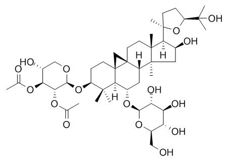
Astragaloside I
Astragaloside Ι may protect the cerebral tissue against the free radical damage in ischemia and inhibit the activation of BV-2 cells induced by LPS through suppressing the activation of PI3K/Akt/NF-κB pathway. It stimulates osteoblast differentiation through the Wnt/β-catenin signaling pathway, which also activates the BMP pathway and RANK pathway.
Inquire / Order:
manager@chemfaces.com
Technical Inquiries:
service@chemfaces.com
Tel:
+86-27-84237783
Fax:
+86-27-84254680
Address:
1 Building, No. 83, CheCheng Rd., Wuhan Economic and Technological Development Zone, Wuhan, Hubei 430056, PRC
Providing storage is as stated on the product vial and the vial is kept tightly sealed, the product can be stored for up to
24 months(2-8C).
Wherever possible, you should prepare and use solutions on the same day. However, if you need to make up stock solutions in advance, we recommend that you store the solution as aliquots in tightly sealed vials at -20C. Generally, these will be useable for up to two weeks. Before use, and prior to opening the vial we recommend that you allow your product to equilibrate to room temperature for at least 1 hour.
Need more advice on solubility, usage and handling? Please email to: service@chemfaces.com
The packaging of the product may have turned upside down during transportation, resulting in the natural compounds adhering to the neck or cap of the vial. take the vial out of its packaging and gently shake to let the compounds fall to the bottom of the vial. for liquid products, centrifuge at 200-500 RPM to gather the liquid at the bottom of the vial. try to avoid loss or contamination during handling.
Sci Rep.2017, 7:467-479
Inflammation2015, 38(1):445-55
Phytochemistry.2017, 141:162-170
Biomed Pharmacother.2024, 175:116770.
J Microbiol Immunol Infect.2021, S1684-1182(21)00142-0.
Jour. of Stored Pro & Postharvest Res.2016, 7(3):32-36
Huazhong Agricultural University2022, pp34.
Int J Mol Sci.2018, 19(9):E2528
Oncology Letters2018, 4690-4696
Inflammation.2021, doi: 10.1007
Related and Featured Products
Phytother Res. 2016 Oct;30(10):1680-8.
Astragaloside I Stimulates Osteoblast Differentiation Through the Wnt/β-catenin Signaling Pathway.[Pubmed:
27397144 ]
Astragaloside I (As-I), one of the main active ingredients in Astragalus membranaceus, is believed to have osteogenic properties, but this hypothesis has not been investigated in detail.
METHODS AND RESULTS:
In the present work, the As-I-induced osteogenic effects and its underlying mechanism were studied in MC3T3-E1 cells. The results indicated that the cellular levels of ALP and extracellular matrix calcium increased in a dose-dependent manner by As-I. To clarify the mechanisms involved in this process, the effect of As-I on the key osteogenic-related genes was investigated. We found that As-I stimulated the expression of β-catenin and Runx2 in MC3T3-E1 cells, which play central roles in the Wnt/β-catenin signaling pathway, suggesting that As-I could promote osteoblastic differentiation by regulating the Wnt/β-catenin signaling pathway. Moreover, the osteogenic effect of As-I could be inhibited by DKK-1, which is the classical inhibitor of Wnt/β-catenin-signaling pathway. Furthermore, As-I also increased BMP-2, BGP and OPG/RANKL expression, which are also activated by Wnt/β-catenin signaling pathway.
CONCLUSIONS:
Taken together, our findings show that As-I stimulates osteoblast differentiation through the Wnt/β-catenin signaling pathway, which also activates the BMP pathway and RANK pathway, thus highlighting the As-I for pharmaceutical and medicinal applications such as treating bone disease.
Chin Med J (Engl). 2013;126(8):1551-4.
Protective effects of astragalus extract against intermittent hypoxia-induced hippocampal neurons impairment in rats.[Pubmed:
23595393]
Intermittent hypoxia is the main pathophysiological cause of the obstructive sleep apnea syndrome. Astragalus shows improvement of spatial learning and memory abilities under intermittent hypoxia. Our study aimed to investigate the protective effect of astragalus against intermittent hypoxia induced-hippocampal neurons impairment in rats and lay the theoretical foundation for the sleep apnea improvement in cognitive function by astragalus.
METHODS AND RESULTS:
Male Wistar rats were divided into 4 groups: blank control group, normoxia group, intermittent hypoxia group and astragalus treated intermittent hypoxia group. After 6-week treatment, apoptosis of neurons was evaluated by terminal deoxynucleotidyl-transferase-mediated dUTP nick end-labeling (TUNEL) assay. Furthermore, the expression of HIF-1a was detected by real-time reverse transcription polymerase chain reaction (RT-PCR) at the mRNA level as well as by immunohistochemistry (IHC) and Western blotting at the protein level.
HPLC analysis indicated that Astragaloside IV, Astragaloside II and Astragaloside I were the main compounds in astragals extract. Astragalus extract reduced the apoptosis of hippocampal neurons (P < 0.05) and decreased the expression of HIF-1a at both the mRNA and protein levels in hippocampus compared with non-treated groups (P < 0.05).
CONCLUSIONS:
Astragalus protects against intermittent hypoxia-induced hippocampal neurons impairment in rats.
China Journal of Traditional Chinese Medicine & Pharmacy, 2016(05).
Astragaloside I inhibited the activation of BV-2 cell induced by LPS through modulation of PI3K/Akt/NF-κB pathway[Reference:
WebLink]
To investigate the effect and mechanism of Astragaloside I(AST I) on the activation of microglial cell line BV-2 induced by LPS.
METHODS AND RESULTS:
After pre-treated with AST I(25, 50, 100μmol/L) for 2 h, BV-2 cells were stimulated with LPS(200ng/m L) for 20 h. Thereafter, the cells and the culture medium were collected. Cell viability was measured by CCK-8 method. Concentration of TNF-α was detected by using ELISA kit. Secretion of NO in medium was assayed with Greiss approach. Gene expressions of CD11 b, TNF-α, i NOS, and IL-1β m RNA were detected with quantitative PCR. Protein expression of PI3K/Akt/NF-κB pathway was detected using Western blotting assay. Neuclear translocation of p-NF-κB was monitored with immunocytochemistry. AST I inhibited the secretion of TNF-α and NO on BV-2 cells upon LPS stimulation(P0.001) without affecting cell viability. It could prevent the up-regulation of gene expressions of TNF-α, i NOS and IL-1β(P0.001, P0.001, P0.05) but not that of CD11 b. Meanwhile, AST I suppressed the increase of i NOS and COX-2 protein induced by LPS. Further study disclosed that AST I reduced nuclear translocation of activated NF-κB and deceased phosphorylation of PI3 K, Akt, NF-κB, and IκB.
CONCLUSIONS:
Astragaloside I inhibited the activation of BV-2 cells induced by LPS through suppressing the activation of PI3K/Akt/NF-κB pathway, therefore, reduced the nuclear translocation of phosphorylated NF-κB, leading to the down-regulation of the gene expressions of TNF-α, i NOS and IL-1β and thus lessened the production of i NOS, TNF-α and COX-2 protein.
Chinese Journal of Clinical Neurosciences, 1998(03):146-8.
Effects of Astragaloside I on Cerebral GSH-px and MDA Contents Following Middle Cerebral Artery Occlusion in Mice[Reference:
WebLink]
Free radicals formation and lipid peroxidation were thought to potentiate ischemic brain cell injury.
METHODS AND RESULTS:
In the present study DTNB method and a-thiobarbituric acid method were applied to measure the activity of GSH-px and the concentration of MDA respectively following MCAO in mice. The results showed that there was no significant difference in cerebral GSHpx activity between ischemic group and sham operated group, while MDA concentration markedly increased in ischemic group compared with the sham operated group (P0. 01). Injection of Astragaloside I intraperitoneally at 1 hour before the MCAO mildly enhanced GSH-px activity, but markedly decreased MDA concentration (P0. 01).
CONCLUSIONS:
These facts suggest that Astragaloside I might protect the cerebral tissue against the free radical damage in ischemia.


