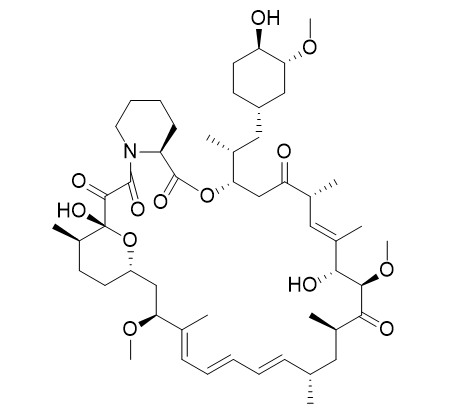
Rapamycin (Sirolimus)
Rapamycin (Sirolimus, AY 22989, NSC-2260804) is a specific mTOR inhibitor with IC50 of ~0.1 nM HEK293 cells.
Inquire / Order:
manager@chemfaces.com
Technical Inquiries:
service@chemfaces.com
Tel:
+86-27-84237783
Fax:
+86-27-84254680
Address:
1 Building, No. 83, CheCheng Rd., Wuhan Economic and Technological Development Zone, Wuhan, Hubei 430056, PRC
Providing storage is as stated on the product vial and the vial is kept tightly sealed, the product can be stored for up to
24 months(2-8C).
Wherever possible, you should prepare and use solutions on the same day. However, if you need to make up stock solutions in advance, we recommend that you store the solution as aliquots in tightly sealed vials at -20C. Generally, these will be useable for up to two weeks. Before use, and prior to opening the vial we recommend that you allow your product to equilibrate to room temperature for at least 1 hour.
Need more advice on solubility, usage and handling? Please email to: service@chemfaces.com
The packaging of the product may have turned upside down during transportation, resulting in the natural compounds adhering to the neck or cap of the vial. take the vial out of its packaging and gently shake to let the compounds fall to the bottom of the vial. for liquid products, centrifuge at 200-500 RPM to gather the liquid at the bottom of the vial. try to avoid loss or contamination during handling.
Asian J Beauty Cosmetol2024, 22(1): 103-112.
Food Chem.2023, 427:136647.
J Applied Biological Chemistry2021, 64(2):185-192
Biol Pharm Bull.2017, 40(6):797-806
Acta Physiologiae Plantarum2016, 38:7
Plant Direct.2021, 5(4):e00318.
Int J Mol Med.2016, 37(2):501-8
Processes2021, 9(1), 153;
Front Pharmacol.2022, 13:806869.
Sci Rep.2023, 13(1):21690.
Related and Featured Products
Nat Med,2002 Feb;8(2):128-35.
Rapamycin inhibits primary and metastatic tumor growth by antiangiogenesis: involvement of vascular endothelial growth factor.[Pubmed:
11821896]
Conventional immunosuppressive drugs have been used effectively to prevent immunologic rejection in organ transplantation. Individuals taking these drugs are at risk, however, for the development and recurrence of cancer.
METHODS AND RESULTS:
In the present study we show that the new immunosuppressive drug rapamycin (RAPA) may reduce the risk of cancer development while simultaneously providing effective immunosuppression. Experimentally, RAPA inhibited metastatic tumor growth and angiogenesis in in vivo mouse models. In addition, normal immunosuppressive doses of RAPA effectively controlled the growth of established tumors. In contrast, the most widely recognized immunosuppressive drug, cyclosporine, promoted tumor growth. From a mechanistic perspective, RAPA showed antiangiogenic activities linked to a decrease in production of vascular endothelial growth factor (VEGF) and to a markedly inhibited response of vascular endothelial cells to stimulation by VEGF.
CONCLUSIONS:
Thus, the use of RAPA, instead of cyclosporine, may reduce the chance of recurrent or de novo cancer in high-risk transplant patients.
J Biol Chem, 2007 May 4;282(18):13395-401.
The rapamycin-binding domain of the protein kinase mammalian target of rapamycin is a destabilizing domain.[Pubmed:
17350953]
Rapamycin is an immunosuppressive drug that binds simultaneously to the 12-kDa FK506- and rapamycin-binding protein (FKBP12, or FKBP) and the FKBP-rapamycin binding (FRB) domain of the mammalian target of rapamycin (mTOR) kinase. The resulting ternary complex has been used to conditionally perturb protein function, and one such method involves perturbation of a protein of interest through its mislocalization.
METHODS AND RESULTS:
We synthesized two rapamycin derivatives that possess large substituents at the C-16 position within the FRB-binding interface, and these derivatives were screened against a library of FRB mutants using a three-hybrid assay in Saccharomyces cerevisiae. Several FRB mutants responded to one of the rapamycin derivatives, and twenty of these mutants were further characterized in mammalian cells. The mutants most responsive to the ligand were fused to yellow fluorescent protein, and fluorescence levels in the presence and absence of the ligand were measured to determine stability of the fusion proteins. Wild-type and mutant FRB domains were expressed at low levels in the absence of the rapamycin derivative, and expression levels rose up to 10-fold upon treatment with ligand. The synthetic rapamycin derivatives were further analyzed using quantitative mass spectrometry, and one of the compounds was found to contain contaminating rapamycin. Furthermore, uncontaminated analogs retained the ability to inhibit mTOR, although with diminished potency relative to rapamycin.
CONCLUSIONS:
The ligand-dependent stability displayed by wild-type FRB and FRB mutants as well as the inhibitory potential and purity of the rapamycin derivatives should be considered as potentially confounding experimental variables when using these systems.
Cancer Res,2005 Apr 15;65(8):3336-46.
Synergistic augmentation of rapamycin-induced autophagy in malignant glioma cells by phosphatidylinositol 3-kinase/protein kinase B inhibitors.[Pubmed:
15833867]
Cell lines:U87-MG, T98G, and U373-MG
Concentrations: Dissolved in DMSO, final concentrations ~25 μM
Incubation Time: 72 hours
Method:
Cells are exposed to various concentrations of Rapamycin for 72 hours. For the assessment of cell viability, cells are collected by trypsinization, stained with trypan blue, and the viable cells in each well are counted. For the determination of cell cycle, cells are trypsinized, fixed with 70% ethanol, and stained with propidium iodide using a flow cytometry reagent set. Samples are analyzed for DNA content using a FACScan flow cytometer and CellQuest software. For apoptosis detection, cells are stained with the terminal deoxynucleotidyl transferase-mediated dUTP nick end labeling (TUNEL) technique using an ApopTag apoptosis detection kit. To detect the development of acidic vesicular organelles (AVO), cells are stained with acridine orange (1 μg/mL) for 15 minutes, and examined under a fluorescence microscope. To quantify the development of AVOs, cells are stained with acridine orange (1 μg/mL) for 15 minutes, removed from the plate with trypsin-EDTA, and analyzed using the FACScan flow cytometer and CellQuest software. To analyze the autophagic process, cells are incubated for 10 minutes with 0.05 mM monodansylcadaverine at 37 °C and are then observed under a fluorescence microscope.
Cancer Cell,2006 Aug;10(2):159-70.
Pathological angiogenesis is induced by sustained Akt signaling and inhibited by rapamycin.[Pubmed:
16904613]
Animal Models:Athymic Nu/Nu mice inoculated subcutaneously with VEGF-A-expressing C6 rat glioma cells
Dosages: ~4 mg/kg/day
Administration: Injection i.p.


