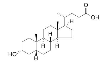
Lithocholic acid
Lithocholic acid is a toxic secondary bile acid, causes intrahepatic cholestasis, has tumor-promoting activity, its toxic effect can be protected after it activates the vitamin D receptor, PXR and FXR.Lithocholic acid is a vitamin D receptor (VDR) ligand, it can activate the VDR to block inflammatory signals in colon cells.
Inquire / Order:
manager@chemfaces.com
Technical Inquiries:
service@chemfaces.com
Tel:
+86-27-84237783
Fax:
+86-27-84254680
Address:
1 Building, No. 83, CheCheng Rd., Wuhan Economic and Technological Development Zone, Wuhan, Hubei 430056, PRC
Providing storage is as stated on the product vial and the vial is kept tightly sealed, the product can be stored for up to
24 months(2-8C).
Wherever possible, you should prepare and use solutions on the same day. However, if you need to make up stock solutions in advance, we recommend that you store the solution as aliquots in tightly sealed vials at -20C. Generally, these will be useable for up to two weeks. Before use, and prior to opening the vial we recommend that you allow your product to equilibrate to room temperature for at least 1 hour.
Need more advice on solubility, usage and handling? Please email to: service@chemfaces.com
The packaging of the product may have turned upside down during transportation, resulting in the natural compounds adhering to the neck or cap of the vial. take the vial out of its packaging and gently shake to let the compounds fall to the bottom of the vial. for liquid products, centrifuge at 200-500 RPM to gather the liquid at the bottom of the vial. try to avoid loss or contamination during handling.
Biomed Pharmacother.2022, 145:112474.
Sci Rep.2015, 5:13194
J Biomol Struct Dyn.2023, 1-21.
Analytical sci. & Tech2016, 186-193
Food Chem.2021, 360:130063.
Int J Pharm.2022, 618:121636.
Phytomedicine.2019, 62:152962
Curr Issues Mol Biol.2023, 45(2):1587-1600.
J Med Food.2021, 24(2):151-160.
Food Bioscience2023, 53:102687
Related and Featured Products
Jpn J Cancer Res. 1998 Nov;89(11):1154-9.
Lithocholic acid, a putative tumor promoter, inhibits mammalian DNA polymerase beta.[Pubmed:
9914784]
Lithocholic acid (LCA), one of the major components in secondary bile acids, promotes carcinogenesis in rat colon epithelial cells induced by N-methyl-N'-nitro-N-nitrosoguanidine (MNNG), which methylates DNA. Base-excision repair of DNA lesions caused by the DNA methylating agents requires DNA polymerase beta (pol beta).
METHODS AND RESULTS:
In the present study, we examined 17 kinds of bile acids with respect to inhibition of mammalian DNA polymerases in vitro. Among them, only LCA and its derivatives inhibited DNA polymerases, while other bile acids were not inhibitory. Among eukaryotic DNA polymerases alpha, beta, delta, epsilon, and gamma, pol beta was the most sensitive to inhibition by LCA. The inhibition mode of pol beta was non-competitive with respect to the DNA template-primer and was competitive with the substrate, dTTP, with the Ki value of 10 microM. Chemical structures at the C-7 and C-12 positions in the sterol skeleton are important for the inhibitory activity of LCA.
CONCLUSIONS:
This inhibition could contribute to the tumor-promoting activity of LCA.
Ann Clin Biochem. 2009 Jan;46(Pt 1):44-9.
Lithocholic acid as a biomarker of intrahepatic cholestasis of pregnancy during ursodeoxycholic acid treatment.[Pubmed:
19103957 ]
The diagnosis and treatment of intrahepatic cholestasis of pregnancy (ICP) has important implications on fetal health. The biochemical parameter commonly used in the diagnosis of ICP is the determination of the concentration of total serum bile acids (TSBA). However, bile acid profile, especially Lithocholic acid (LCA) analysis is a more sensitive and specific biomarker for differential diagnosis of this pathology and also could be an alternative to evaluate the efficiency of ursodeoxycholic acid (UDCA) for ICP treatment.
METHODS AND RESULTS:
Serum bile acid (SBA) profiles including LCA determination, were studied in 28 ICP patients using a capillary electrophoresis method. The effects of UDCA treatment on bile acid profile, were analysed in 23 out of 28 ICP patients and the two samples obtained before and 15 days after treatment were compared. Two samples taken as controls were also obtained from each of five patients without therapy.
A dramatic decrease in LCA concentrations and maintenance of TSBA concentrations were found in all patients after UDCA therapy, whereas SBA profiles together with LCA values did not change in patients without therapy.
CONCLUSIONS:
We propose LCA as an alternative biomarker and a more sensitive parameter than TSBA to evaluate the effectiveness of UDCA treatment, at least in ICP patients from Argentina.
Journal of Biological Chemistry,2002, 277(35) :31441-7.
Lithocholic acid decreases expression of bile salt export pump through farnesoid X receptor antagonist activity[Reference:
WebLink]
Bile salt export pump (BSEP) is a major bile acid transporter in the liver. Mutations in BSEP result in progressive intrahepatic cholestasis, a severe liver disease that impairs bile flow and causes irreversible liver damage. BSEP is a target for inhibition and down-regulation by drugs and abnormal bile salt metabolites, and such inhibition and down-regulation may result in bile acid retention and intrahepatic cholestasis.
METHODS AND RESULTS:
In this study, we quantitatively analyzed the regulation of BSEP expression by FXR ligands in primary human hepatocytes and HepG2 cells. We demonstrate that BSEP expression is dramatically regulated by ligands of the nuclear receptor farnesoid X receptor (FXR). Both the endogenous FXR agonist chenodeoxycholate (CDCA) and synthetic FXR ligand GW4064 effectively increased BSEP mRNA in both cell types. This up-regulation was readily detectable at as early as 3 h, and the ligand potency for BSEP regulation correlates with the intrinsic activity on FXR. These results suggest BSEP as a direct target of FXR and support the recent report that the BSEP promoter is transactivated by FXR. In contrast to CDCA and GW4064, lithocholate (LCA), a hydrophobic bile acid and a potent inducer of cholestasis, strongly decreased BSEP expression. Previous studies did not identify LCA as an FXR antagonist ligand in cells, but we show here that LCA is an FXR antagonist with partial agonist activity in cells. In an in vitro co-activator association assay, LCA decreased CDCA- and GW4064-induced FXR activation with an IC50 of 1 μM. In HepG2 cells, LCA also effectively antagonized GW4064-enhanced FXR transactivation. These data suggest that the toxic and cholestatic effect of LCA in animals may result from its down-regulation of BSEP through FXR.
CONCLUSIONS:
Taken together, these observations indicate that FXR plays an important role in BSEP gene expression and that FXR ligands may be potential therapeutic drugs for intrahepatic cholestasis.
J Steroid Biochem Mol Biol. 2008 Jul;111(1-2):37-40.
Lithocholic acid down-regulation of NF-kappaB activity through vitamin D receptor in colonic cancer cells.[Pubmed:
18515093 ]
Lithocholic acid (LCA), a secondary bile acid, is a vitamin D receptor (VDR) ligand. 1,25-Dihydroxyvitamin D(3) (1,25(OH)(2)D(3)), the hormonal form of vitamin D, is involved in the anti-inflammatory action through VDR. Therefore, we hypothesize that LCA acts like 1,25(OH)(2)D(3) to drive anti-inflammatory signals.
METHODS AND RESULTS:
In present study, we used human colonic cancer cells to assess the role of LCA in regulation of the pro-inflammatory NF-kappaB pathway. We found that LCA treatment increased VDR levels, mimicking the effect of 1,25(OH)(2)D(3). LCA pretreatment inhibited the IL-1beta-induced IkappaBalpha degradation and decreased the NF-kappaB p65 phosphorylation. We also measured the production of IL-8, a well-known NF-kappaB target gene, as a read-out of the biological effect of LCA expression on NF-kappaB pathway. LCA significantly decreased IL-8 secretion induced by IL-1beta. These LCA-induced effects were very similar to those of 1,25(OH)(2)D(3.) Thus, LCA recapitulated the effects of 1,25(OH)(2)D(3) on IL-1beta stimulated cells. Mouse embryonic fibroblast (MEF) cells lacking VDR have intrinsically high NF-kappaB activity. LCA pretreatment was not able to prevent TNFalpha-induced IkappaBalpha degradation in MEF VDR (-/-), whereas LCA stabilized IkappaBalpha in MEF VDR (+/-) cells.
CONCLUSIONS:
Collectively, our data indicated that LCA activated the VDR to block inflammatory signals in colon cells.


