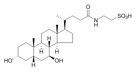
Tauroursodeoxycholic acid
Tauroursodeoxycholic acid (TUDCA) may enhance the secretory capacity of cholestatic hepatocytes by stimulation of exocytosis and insertion of transport proteins into apical membranes via PKC-dependent mechanisms. TUDCA acts as a chemical chaperone to enhance protein folding and ameliorate ER stress, increases insulin sensitivity, it may be an effective pharmacological approach for treating insulin resistance. TUDCA has wide-range neuroprotective effects, it is a strong modulator of AbetaE22Q-triggered apoptosis, it may provide a potentially useful treatment in patients with hemorrhagic stroke and perhaps other acute brain injuries associated with cell death by apoptosis.
Inquire / Order:
manager@chemfaces.com
Technical Inquiries:
service@chemfaces.com
Tel:
+86-27-84237783
Fax:
+86-27-84254680
Address:
1 Building, No. 83, CheCheng Rd., Wuhan Economic and Technological Development Zone, Wuhan, Hubei 430056, PRC
Providing storage is as stated on the product vial and the vial is kept tightly sealed, the product can be stored for up to
24 months(2-8C).
Wherever possible, you should prepare and use solutions on the same day. However, if you need to make up stock solutions in advance, we recommend that you store the solution as aliquots in tightly sealed vials at -20C. Generally, these will be useable for up to two weeks. Before use, and prior to opening the vial we recommend that you allow your product to equilibrate to room temperature for at least 1 hour.
Need more advice on solubility, usage and handling? Please email to: service@chemfaces.com
The packaging of the product may have turned upside down during transportation, resulting in the natural compounds adhering to the neck or cap of the vial. take the vial out of its packaging and gently shake to let the compounds fall to the bottom of the vial. for liquid products, centrifuge at 200-500 RPM to gather the liquid at the bottom of the vial. try to avoid loss or contamination during handling.
Srinagarind Medical Journal2019, 34(1)
Int J Mol Sci.2021, 22(21):11447.
Food Funct.2022, D1FO03838A.
J. Traditional Thai Medical Res. 2022,8(1):1-14.
Plant J.2021, 107(6):1711-1723.
Front Pharmacol.2022, 13:806869.
Int J Vet Sci Med.2024, 12(1):134-147.
Methods Protoc.2024, 7(6):95.
Agronomy2020, 10(3),388.
PLoS One.2022, 17(4):e0267007.
Related and Featured Products
Diabetes,2010, 59(8):1899-1905.
Tauroursodeoxycholic Acid may improve liver and muscle but not adipose tissue insulin sensitivity in obese men and women.[Pubmed:
20522594 ]
Insulin resistance is commonly associated with obesity. Studies conducted in obese mouse models found that endoplasmic reticulum (ER) stress contributes to insulin resistance, and treatment with Tauroursodeoxycholic acid (TUDCA), a bile acid derivative that acts as a chemical chaperone to enhance protein folding and ameliorate ER stress, increases insulin sensitivity. The purpose of this study was to determine the effect of TUDCA therapy on multiorgan insulin action and metabolic factors associated with insulin resistance in obese men and women.
METHODS AND RESULTS:
Twenty obese subjects ([means +/- SD] aged 48 +/- 11 years, BMI 37 +/- 4 kg/m2) were randomized to 4 weeks of treatment with TUDCA (1,750 mg/day) or placebo. A two-stage hyperinsulinemic-euglycemic clamp procedure in conjunction with stable isotopically labeled tracer infusions and muscle and adipose tissue biopsies were used to evaluate in vivo insulin sensitivity, cellular factors involved in insulin signaling, and cellular markers of ER stress. RESULTS Hepatic and muscle insulin sensitivity increased by approximately 30% (P < 0.05) after treatment with TUDCA but did not change after placebo therapy. In addition, therapy with TUDCA, but not placebo, increased muscle insulin signaling (phosphorylated insulin receptor substrate(Tyr) and Akt(Ser473) levels) (P < 0.05). Markers of ER stress in muscle or adipose tissue did not change after treatment with either TUDCA or placebo.
CONCLUSIONS:
These data demonstrate that TUDCA might be an effective pharmacological approach for treating insulin resistance. Additional studies are needed to evaluate the target cells and mechanisms responsible for this effect.
Gut. 2008 Oct;57(10):1448-54.
Tauroursodeoxycholic acid exerts anticholestatic effects by a cooperative cPKC alpha-/PKA-dependent mechanism in rat liver[Pubmed:
18583398 ]
Ursodeoxycholic acid (UDCA) exerts anticholestatic effects in part by protein kinase C (PKC)-dependent mechanisms. Its taurine conjugate, TUDCA, is a cPKC alpha agonist. We tested whether protein kinase A (PKA) might contribute to the anticholestatic action of TUDCA via cooperative cPKC alpha-/PKA-dependent mechanisms in taurolithocholic acid (TLCA)-induced cholestasis.
METHODS AND RESULTS:
In perfused rat liver, bile flow was determined gravimetrically, organic anion secretion spectrophotometrically, lactate dehydrogenase (LDH) release enzymatically, cAMP response-element binding protein (CREB) phosphorylation by immunoblotting, and cAMP by immunoassay. PKC/PKA inhibitors were tested radiochemically. In vitro phosphorylation of the conjugate export pump, Mrp2/Abcc2, was studied in rat hepatocytes and human Hep-G2 hepatoma cells.
In livers treated with TLCA (10 micromol/l)+TUDCA (25 micromol/l), combined inhibition of cPKC by the cPKC-selective inhibitor Gö6976 (100 nmol/l) or the non-selective PKC inhibitor staurosporine (10 nmol/l) and of PKA by H89 (100 nmol/l) reduced bile flow by 36% (p<0.05) and 48% (p<0.01), and secretion of the Mrp2/Abcc2 substrate, 2,4-dinitrophenyl-S-glutathione, by 31% (p<0.05) and 41% (p<0.01), respectively; bile flow was unaffected in control livers or livers treated with TUDCA only or TLCA+taurocholic acid. Inhibition of cPKC or PKA alone did not affect the anticholestatic action of TUDCA. Hepatic cAMP levels and CREB phosphorylation as readout of PKA activity were unaffected by the bile acids tested, suggesting a permissive effect of PKA for the anticholestatic action of TUDCA. Rat and human hepatocellular Mrp2 were phosphorylated by phorbol ester pretreatment and recombinant cPKC alpha, nPKC epsilon, and PKA, respectively, in a staurosporine-sensitive manner.
CONCLUSIONS:
UDCA conjugates exert their anticholestatic action in bile acid-induced cholestasis in part via cooperative post-translational cPKC alpha-/PKA-dependent mechanisms. Hepatocellular Mrp2 may be one target of bile acid-induced kinase activation.
Hepatology. 2001 May;33(5):1206-16.
Tauroursodeoxycholic acid inserts the apical conjugate export pump, Mrp2, into canalicular membranes and stimulates organic anion secretion by protein kinase C-dependent mechanisms in cholestatic rat liver.[Pubmed:
11343250 ]
Ursodeoxycholic acid (UDCA) exerts anticholestatic effects by undefined mechanisms. Previous work suggested that UDCA stimulates biliary exocytosis via Ca(++)- and protein kinase C (PKC)-dependent mechanisms. Therefore, the effect of taurine-conjugated UDCA (TUDCA) was studied in the experimental model of taurolithocholic acid (TLCA)-induced cholestasis on bile flow, hepatobiliary exocytosis, distribution of PKC isoforms, and density of the apical conjugate export pump, Mrp2, in canalicular membranes.
METHODS AND RESULTS:
Isolated perfused rat livers were preloaded with horseradish peroxidase (HRP), a marker of vesicular exocytosis, and were perfused with bile acids or dimethylsulfoxide (control) only. PKC isoform distribution and membrane density of Mrp2 were studied using immunoblotting and immunoelectron-microscopic techniques. Biliary secretion of the Mrp2 substrate, 2,4-dinitrophenyl-S-glutathione (GS-DNP), was studied in the presence or absence of the PKC inhibitor, bisindolylmaleimide I (BIM-I; 1 micromol/L). TLCA (10 micromol/L) impaired bile flow by 51%; biliary secretion of HRP and GS-DNP by 46% and 95%, respectively; membrane binding of the Ca(++)-sensitive alpha-isoform of PKC by 32%; and density of Mrp2 in the canalicular membrane by 79%. TUDCA (25 micromol/L) reversed the effects of TLCA on bile flow, secretion of HRP and GS-DNP, and distribution of alpha-PKC. TUDCA reduced membrane binding of epsilon-PKC and increased Mrp2 density 4-fold in canalicular membranes of cholestatic hepatocytes. BIM-I inhibited the effect of TUDCA on GS-DNP secretion in cholestatic livers by 49% without affecting secretion in controls.
CONCLUSIONS:
In conclusion, TUDCA may enhance the secretory capacity of cholestatic hepatocytes by stimulation of exocytosis and insertion of transport proteins into apical membranes via PKC-dependent mechanisms.
Cell Mol Life Sci. 2009 Mar;66(6):1094-104.
Tauroursodeoxycholic acid prevents E22Q Alzheimer's Abeta toxicity in human cerebral endothelial cells.[Pubmed:
19189048 ]
The vasculotropic E22Q mutant of the amyloid-beta (Abeta) peptide is associated with hereditary cerebral hemorrhage with amyloidosis Dutch type. The cellular mechanism(s) of toxicity and nature of the AbetaE22Q toxic assemblies are not completely understood.
METHODS AND RESULTS:
Comparative assessment of structural parameters and cell death mechanisms elicited in primary human cerebral endothelial cells by AbetaE22Q and wild-type Abeta revealed that only AbetaE22Q triggered the Bax mitochondrial pathway of apoptosis. AbetaE22Q neither matched the fast oligomerization kinetics of Abeta42 nor reached its predominant beta-sheet structure, achieving a modest degree of oligomerization with a secondary structure that remained a mixture of beta and random conformations. The endogenous molecule Tauroursodeoxycholic acid (TUDCA) was a strong modulator of AbetaE22Q-triggered apoptosis but did not significantly change the secondary structures and fibrillogenic propensities of Abeta peptides.
CONCLUSIONS:
These data dissociate the pro-apoptotic properties of Abeta peptides from their distinct mechanisms of aggregation/fibrillization in vitro, providing new perspectives for modulation of amyloid toxicity.
Proc Natl Acad Sci U S A. 2003 May 13;100(10):6087-92.
Tauroursodeoxycholic acid reduces apoptosis and protects against neurological injury after acute hemorrhagic stroke in rats.[Pubmed:
12721362]
Tauroursodeoxycholic acid (TUDCA), an endogenous bile acid, modulates cell death by interrupting classic pathways of apoptosis. Intracerebral hemorrhage (ICH) is a devastating acute neurological disorder, without effective treatment, in which a significant loss of neuronal cells is thought to occur by apoptosis.
METHODS AND RESULTS:
In this study, we evaluated whether TUDCA can reduce brain injury and improve neurological function after ICH in rats. Administration of TUDCA before or up to 6 h after stereotaxic collagenase injection into the striatum reduced lesion volumes at 2 days by as much as 50%. Apoptosis was approximately 50% decreased in the area immediately surrounding the hematoma and was associated with a similar inhibition of caspase activity. These changes were also associated with improved neurobehavioral deficits as assessed by rotational asymmetry, limb placement, and stepping ability. Furthermore, TUDCA treatment modulated expression of certain Bcl-2 family members, as well as NF-kappaB activity. In addition to its protective action at the mitochondrial membrane, TUDCA also activated the Akt-1protein kinase Balpha survival pathway and induced Bad phosphorylation at Ser-136.
CONCLUSIONS:
In conclusion, reduction of brain injury underlies the wide-range neuroprotective effects of TUDCA after ICH. Thus, given its clinical safety, TUDCA may provide a potentially useful treatment in patients with hemorrhagic stroke and perhaps other acute brain injuries associated with cell death by apoptosis.


