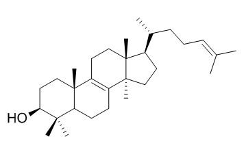
Lanosterol
Lanosterol is a key triterpenoid intermediate in the biosynthesis of Cholesterol. Lanosterol induces mild depolarization of mitochondria and promotes autophagy, it is indicative of altered Lanosterol metabolism during PD pathogenesis. 10 μM Lanosterol during IVM in medium without serum and bases on recombinant human chorionic gonadotrophin has a positive effect on maturation of prepubertal ewe oocytes.
Inquire / Order:
manager@chemfaces.com
Technical Inquiries:
service@chemfaces.com
Tel:
+86-27-84237783
Fax:
+86-27-84254680
Address:
1 Building, No. 83, CheCheng Rd., Wuhan Economic and Technological Development Zone, Wuhan, Hubei 430056, PRC
Providing storage is as stated on the product vial and the vial is kept tightly sealed, the product can be stored for up to
24 months(2-8C).
Wherever possible, you should prepare and use solutions on the same day. However, if you need to make up stock solutions in advance, we recommend that you store the solution as aliquots in tightly sealed vials at -20C. Generally, these will be useable for up to two weeks. Before use, and prior to opening the vial we recommend that you allow your product to equilibrate to room temperature for at least 1 hour.
Need more advice on solubility, usage and handling? Please email to: service@chemfaces.com
The packaging of the product may have turned upside down during transportation, resulting in the natural compounds adhering to the neck or cap of the vial. take the vial out of its packaging and gently shake to let the compounds fall to the bottom of the vial. for liquid products, centrifuge at 200-500 RPM to gather the liquid at the bottom of the vial. try to avoid loss or contamination during handling.
Biomol Ther (Seoul).2023, 31(1):40-47.
Biology (Basel).2020, 9(11):363.
Sci Rep. 2017, 12953(7)
Pharmaceuticals (Basel).2024, 17(3):341.
Evid Based Complement Alternat Med.2018, 2018:3610494
J Liq Chromatogr R T2018, 41(12):761-769
Phytother Res.2022, 10.1002:ptr.7592.
Pol J Microbiol.2021, 70(1):117-130.
World J Mens Health.2019, 10.5534
ACS Omega.2023, 9(1):1278-1286.
Related and Featured Products
Zygote. 2014 Feb;22(1):50-7.
Effect of lanosterol on the in vitro maturation in semi-defined culture system of prepubertal ewe oocytes.[Pubmed:
21838962]
The choice of medium and supplements can affect meiotic regulation and may have an impact on the regulation of mammalian oocyte growth and embryonic cell function. The aim of the present study was to assess the effects of oxygen concentration and endogenous Lanosterol on the in vitro maturation (IVM) media without serum and based on recombinant human chorionic gonadotrophin in prepubertal ewe oocytes.
METHODS AND RESULTS:
Firstly, the effect of varying oxygen concentrations (5% and 20%) during IVM in TCM-199 supplemented (4 mg/ml bovine serum albumin (BSA), 100 μM cysteamine, 0.3 mM sodium pyruvate, 0.1 UI/ml recombinant follicle-stimulating hormone (r-FSH; Gonal-F® 75 UI, Serono, Italy), 0.1 UI/ml recombinant leuteinizing hormone (r-LH; Lhadi® 75 UI, Serono, Italy) and 1 μg/ml estradiol-17β) on subsequent nuclear maturation of oocytes examined under ultraviolet light following staining with bisbenzimide (Hoechst 33342) was investigated. Secondly, two concentrations of Lanosterol (0, 10 and 50 μM) were added to the IVM medium. Nuclear maturation of oocytes was examined as previously. Lipid content in oocytes, an important indicator of cytoplasmic maturity, was also measured using Nile red fluorescent stain. The results showed that low oxygen concentration affected the nuclear maturation. Similarly, a significantly higher rate of meiosis resumption was observed with 10 μM (72.3%) of Lanosterol compared with the control (51.8%) or 50 μM of Lanosterol (59.4%). A significantly higher content of lipids was also observed with 10 and 50 μM of Lanosterol (7.3 ± 0.2 × 10(6) and 7.4 ± 0.2 × 10(6) arbitrary units of fluorescence) compared with the control (6.7 ± 0.2 × 10(6) arbitrary units of fluorescence).
CONCLUSIONS:
The results indicate that 10 μM Lanosterol during IVM in medium without serum and based on recombinant human chorionic gonadotrophin has a positive effect on maturation of prepubertal ewe oocytes.
Cell Death Differ. 2012 Mar;19(3):416-27.
Lanosterol induces mitochondrial uncoupling and protects dopaminergic neurons from cell death in a model for Parkinson's disease.[Pubmed:
21818119]
To investigate the anti-HBV constituents in the roots of Euphorbia fischeriana.
METHODS AND RESULTS:
The compounds were isolated by various chromatographic methods and identified by spectroscopic analysis. Some compounds were tested for the anti-HBV activity. Eleven compounds were isolated and identified as tirucalla-5,24-dien-3-ol (1), 24-methyltirucalla-5, 24-dien-3-ol (2), euphol (3), Butyrospermol (4), 24-methylenecycloartenol (5), cycloartenol (6), jolkinolid E (7) helioscopinolide A (8), isoscopoletion (9), dephnoretin (10), and 3, 3'-di-O-methylellagic acid 4'-O-beta-D-xylopyranoside (11).
CONCLUSIONS:
Compounds 1, 2 and 10 were isolated from the genus Euphorbia for the first time. Compounds 3, 4 and 11 were isolated from this species for the first time. Compounds 1, 8, 9 and 11 showed weak anti-HBsAg and anti-HBeAg activity, while compound 10 showed weak anti-HBsAg activity.
Lanosterol Suppresses the Aggregation and Cytotoxicity of Misfolded Proteins Linked with Neurodegenerative Diseases
Lanosterol Suppresses the Aggregation and Cytotoxicity of Misfolded Proteins Linked with Neurodegenerative Diseases[Pubmed:
28102469]
Abstract
Accumulation of misfolded or aberrant proteins in neuronal cells is linked with neurodegeneration and other pathologies. Which molecular mechanisms fail and cause inappropriate folding of proteins and what is their relationship to cellular toxicity is not well known. How does it happen and what are the probable therapeutic or molecular approaches to counter them are also not clear. Here, we demonstrate that treatment of Lanosterol diminishes aberrant proteotoxic aggregation and mitigates their cytotoxicity via induced expression of co-chaperone CHIP and elevated autophagy. The addition of Lanosterol not only reduces aggregation of mutant bonafide misfolded proteins but also effectively prevents accumulation of various mutant disease-prone proteotoxic proteins. Finally, we observed that Lanosterol mitigates cytotoxicity in cells, mediated by different stress-inducing agents. Taken together, our present results suggest that upregulation of cellular molecular chaperones, primarily using small molecules, can probably offer an efficient therapeutic approach in the future against misfolding of different disease-causing proteins and neurodegenerative disorders. Graphical Abstract ᅟ.
Keywords: CHIP; Cell death; Lanosterol; Misfolded proteins; Neurodegeneration.
Theriogenology . 2016 Mar 1;85(4):575-84.
Lanosterol influences cytoplasmic maturation of pig oocytes in vitro and improves preimplantation development of cloned embryos[Pubmed:
26494176]
Abstract
Lanosterol is a precursor of meiosis-activating sterols in the cholesterol biosynthetic pathway and induces a physiological signal that instructs the oocyte to reinitiate meiosis. In this study, we examined the effect of Lanosterol on IVM of porcine oocytes, specifically on nuclear maturation, cytoplasmic maturation by investigating intracellular glutathione (GSH) levels and lipid content, embryonic development after parthenogenetic activation and somatic cell nuclear transfer (SCNT), and on gene expression in cumulus cells, oocytes, and SCNT-derived blastocysts. There was no significant difference in nuclear maturation rates between the control and treatment groups (10, 50, and 100 μM of Lanosterol added to IVM culture medium). Supplementation with 50-μM Lanosterol significantly increased lipid content and GSH levels and decreased reactive oxygen species levels compared with the control. In addition, oocytes treated with 50 μM of Lanosterol exhibited significantly increased blastocyst formation rates and total cell numbers after parthenogenetic activation (30.3% and 63.9 vs. 21.6% and 36.5, respectively) and SCNT (18.2% and 53.7 vs. 12.6% and 37.5, respectively), when compared with the control group. Cumulus cells treated with 50 μM of Lanosterol showed significantly increased 14α-demethylase, Δ14-reductase, and Δ7-reductase mRNA transcript levels. Significantly increased PPARγ, SREBF1, GPX1, and Bcl-2 and decreased Bax transcript levels were observed in mature oocytes treated with 50 μM of Lanosterol compared with the control. SCNT blastocysts derived from 50-μM Lanosterol-treated oocytes had significantly higher POU5F1, FGFR2, and Bcl-2 transcript levels than control SCNT-derived blastocysts. In conclusion, supplementation with 50 μM of Lanosterol during IVM improves preimplantation development of SCNT embryos by elevating lipid content of oocytes, increasing GSH levels, decreasing reactive oxygen species levels, and regulating genes related to the cholesterol biosynthetic pathway in cumulus cells, to lipid metabolism and apoptosis in oocytes, and their developmental potential and apoptosis in blastocysts.
Keywords: Embryonic development; IVM; Lanosterol; Porcine oocyte; Somatic cell nuclear transfer.


