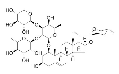
Ophiopogonin D
Ophiopogonin D plays a protective role as an effective antioxidant in H2O2-induced endothelial injury, it can attenuate doxorubicin-induced autophagic cell death by relieving mitochondrial damage in vitro and in vivo, it can be therefore developed as a novel drug for the therapy of cardiovascular disorders. Ophiopogonin D and spicatoside A can increase mucin production and secretion, by directly acting on airway epithelial cells, they could be as expectorants in diverse inflammatory pulmonary diseases. Ophiopogonin D inhibits MCF-7 cell growth via the induction of cell cycle arrest at the G2/M phase.
Inquire / Order:
manager@chemfaces.com
Technical Inquiries:
service@chemfaces.com
Tel:
+86-27-84237783
Fax:
+86-27-84254680
Address:
1 Building, No. 83, CheCheng Rd., Wuhan Economic and Technological Development Zone, Wuhan, Hubei 430056, PRC
Providing storage is as stated on the product vial and the vial is kept tightly sealed, the product can be stored for up to
24 months(2-8C).
Wherever possible, you should prepare and use solutions on the same day. However, if you need to make up stock solutions in advance, we recommend that you store the solution as aliquots in tightly sealed vials at -20C. Generally, these will be useable for up to two weeks. Before use, and prior to opening the vial we recommend that you allow your product to equilibrate to room temperature for at least 1 hour.
Need more advice on solubility, usage and handling? Please email to: service@chemfaces.com
The packaging of the product may have turned upside down during transportation, resulting in the natural compounds adhering to the neck or cap of the vial. take the vial out of its packaging and gently shake to let the compounds fall to the bottom of the vial. for liquid products, centrifuge at 200-500 RPM to gather the liquid at the bottom of the vial. try to avoid loss or contamination during handling.
Int J Mol Sci.2024, 25(5):2914.
Integr Cancer Ther.2018, 17(3):832-843
Babol University of Medical Sciences2024, rs-4289336
Sci Rep.2024, 14(1):31213.
Molecules.2021, 26(6):1635.
ARPN Journal of Eng.& Applied Sci.2016, 2199-2204
Food Control2022, 132:108434.
Int J Mol Sci.2024, 25(20):11227.
Journal of Functional Foods2022, 91:105019.
Plant Cell,Tissue & Organ Culture2016, 127(1):115-121
Related and Featured Products
Mol Med Rep. 2015 Jul;12(1):1493-8.
Ophiopogonin-D suppresses MDA-MB-435 cell adhesion and invasion by inhibiting matrix metalloproteinase-9.[Pubmed:
25816153]
Ophiopogonin D is one of steroidal saponins isolated from the root of the Chinese medicinal plant Ophiopogon japonicas. It has been claimed to possess anti-inflammatory and anti-oxidant properties.
The present study was the first to examine the anti-tumor metastasis properties of Ophiopogonin D.
METHODS AND RESULTS:
An MTT assay showed that Ophiopogonin D inhibited the proliferation of MDA-MB-435 melanoma cells, and decreased invasion was demonstrated using a Transwell invasion assay. Furthermore, adhesion of MDA-MB-435 cells to human umbilical vascular endothelial cells and to fibronectin was inhibited by Ophiopogonin D. Gelatin zymography and western blot analysis showed that Ophiopogonin D inhibited the expression and secretion of matrix metalloproteinase-9 (MMP-9), but not that of MMP-2. Inhibition of phosphorylation of p38 by Ophiopogonin D indicated its inhibition of the mitogen-activated protein kinase pathway.
CONCLUSIONS:
Overall, the results suggested that Ophiopogonin D may be considered as a candidate drug for treating or preventing tumor metastasis.
J Pharmacol Exp Ther. 2015 Jan;352(1):166-74.
Ophiopogonin D attenuates doxorubicin-induced autophagic cell death by relieving mitochondrial damage in vitro and in vivo.[Pubmed:
25378375]
It has been reported that Ophiopogonin D (OP-D), a steroidal glycoside and an active component extracted from Ophiopogon japonicas, promotes antioxidative protection of the cardiovascular system. However, it is unknown whether Ophiopogonin D exerts protective effects against doxorubicin (DOX)-induced autophagic cardiomyocyte injury.
METHODS AND RESULTS:
Here, we demonstrate that DOX induced excessive autophagy through the generation of reactive oxygen species (ROS) in H9c2 cells and in mouse hearts, which was indicated by a significant increase in the number of autophagic vacuoles, LC3-II/LC3-I ratio, and upregulation of the expression of GFP-LC3. Pretreatment with Ophiopogonin D partially attenuated the above phenomena, similar to the effects of treatment with 3-methyladenine. In addition, Ophiopogonin D treatment significantly relieved the disruption of the mitochondrial membrane potential by antioxidative effects through downregulating the expression of both phosphorylated c-Jun N-terminal kinase and extracellular signal-regulated kinase.
CONCLUSIONS:
The ability of Ophiopogonin D to reduce the generation of ROS due to mitochondrial damage and, consequently, to inhibit autophagic activity partially accounts for its protective effects in the hearts against DOX-induced toxicity.
Phytochemistry . 2017 Apr;136:125-132.
Homo-aro-cholestane, furostane and spirostane saponins from the tubers of Ophiopogon japonicus[Pubmed:
28139298]
Abstract
Phytochemical investigation of the tubers of Ophiopogon japonicus led to the isolation of five previously undescribed steroidal saponins, ophiojaponins A-E, together with twelve known ones. The structures of these isolated compounds were elucidated by detailed spectroscopic analyses and chemical methods. Ophiojaponins A-C are rare naturally occurring C29 steroidal glycosides possessing a homo-cholestane skeleton with an aromatized ring E. Ruscogenin 1-O-α-L-rhamnopyranosyl-(1 → 2)-4-O-sulfo-β-D-fucopyranosido-3-O-β-D-glucopyranoside was isolated as single component and its full spectroscopic data was reported for the first time. The isolated steroidal saponins were evaluated for their cytotoxicities against two human tumor cell lines MG-63 and SNU387. Among them, five known spirostane-type glycosides showed cytotoxic activity against both MG-63 and SNU387 cell lines with IC50 values ranging from 0.76 to 27.0 μM.
Keywords: C(27)-steroidal saponins; C(29)-steroidal saponins; Cytotoxic activity; Liliaceae; Ophiopogon japonicus.
BMC Complement Altern Med. 2014 Sep 23;14:350.
A metabonomic study of cardioprotection of ginsenosides, schizandrin, and ophiopogonin D against acute myocardial infarction in rats.[Pubmed:
25249156]
Metabonomics is a useful tool for studying mechanisms of drug treatment using systematic metabolite profiles. Ginsenosides Rg1 and Rb1, Ophiopogonin D, and schizandrin are the main bioactive components of a traditional Chinese formula (Sheng-Mai San) widely used for the treatment of coronary heart disease. It remains unknown the effect of individual bioactive component and how the multi-components in combination affect the treating acute myocardial infarction (AMI).
METHODS AND RESULTS:
Rats were divided into 7 groups and dosed consecutively for 7 days with mono and combined-therapy administrations. Serum samples were analyzed by proton nuclear magnetic resonance (1H NMR) spectroscopy. Partial least squares discriminate analysis (PLS-DA) was employed to distinguish the metabolic profile of rats in different groups and identify potential biomarkers.
Score plots of PLS-DA exhibited that combined-therapy groups were significantly different from AMI group, whereas no differences were observed for mono-therapy groups. We found that AMI caused comprehensive metabolic changes involving stimulation of glycolysis, suppression of fatty acid oxidation, together with disturbed metabolism of arachidonic acid, linoleate, leukotriene, glycerophospholipid, phosphatidylinositol phosphate, and some amino acids. β-hydroxybutyrate, cholines and glucose were regulated by mono-therapy of schizandrin and ginsenosides respectively. Besides these metabolites, combined-therapy ameliorated more of the AMI-induced metabolic changes including glycerol, and O-acetyl glycoprotein. A remarkable reduction of lactate suggested the therapeutic effect of combined-therapy through improving myocardial energy metabolism.
CONCLUSIONS:
This study provided novel metabonomic insights on the mechanism of synergistic cardioprotection of combined-therapy with ginsenosides, schizandrin, and Ophiopogonin D, and demonstrated the potential of discovering new drugs by combining bioactive components from traditional Chinese formula.
Phytomedicine. 2014 Jan 15;21(2):172-6.
Effects of ophiopogonin D and spicatoside A derived from Liriope Tuber on secretion and production of mucin from airway epithelial cells.[Pubmed:
24060215]
In the present study, we investigated whether aqueous extract of Liriope Tuber, Ophiopogonin D and spicatoside A derived from Liriope Tuber affect basal or phorbol ester (phorbol 12-myristate 13-acetate, PMA)-induced airway mucin production and secretion from airway epithelial cells.
METHODS AND RESULTS:
Confluent NCI-H292 cells were treated with each agent for 24 h (basal production) or pretreated with each agent for 30 min and then stimulated with PMA for 24 h (PMA-induced production and secretion), respectively. MUC5AC airway mucin production and secretion were measured by ELISA. The results were as follows: (1) aqueous extract of Liriope Tuber stimulated basal mucin production and did not inhibit but increased PMA-induced mucin production; (2) Ophiopogonin D and spicatoside A stimulated basal mucin production and did not inhibit but increased PMA-induced mucin production; (3) two compounds increased PMA-induced mucin secretion.
CONCLUSIONS:
These results suggest that Ophiopogonin D and spicatoside A can increase mucin production and secretion, by directly acting on airway epithelial cells and, at least in part, explain the traditional use of aqueous extract of Liriope Tuber as expectorants in diverse inflammatory pulmonary diseases.
J Integr Med. 2016 Jan;14(1):51-9.
Ophiopogonin D inhibits cell proliferation, causes cell cycle arrest at G2/M, and induces apoptosis in human breast carcinoma MCF-7 cells.[Pubmed:
26778229]
To investigate the effects of Ophiopogonin D on human breast cancer MCF-7 cells.
METHODS AND RESULTS:
Cell viability was assessed by 3-(4,5-dimethylthiazol-2-yl)-2,5-diphenyltetrazolium bromide and colony formation experiments. Cell cycle was measured with cell cycle flow cytometry and a living cell assay. Apoptosis and terminal deoxynucleoitidyl transferase-mediated dUTP nick end labeling assays were performed to detect the apoptosis of MCF-7 cells induced by Ophiopogonin D. Finally, Western blotting was used to explore the mechanism. Exposure of cells to Ophiopogonin D resulted in marked decreases in viable cells and colony formation with a dose-dependent manner. Treatment of these cells with Ophiopogonin D also resulted in cell cycle arrest at the G(2)/M phase, and increased apoptosis. Mechanistically, Ophiopogonin D-induced G(2)/M cell cycle arrest was associated with down-regulation of cyclin B1. Furthermore, activation of caspase-8 and caspase-9 was involved in Ophiopogonin D-induced apoptosis.
CONCLUSIONS:
The data suggested that Ophiopogonin D inhibits MCF-7 cell growth via the induction of cell cycle arrest at the G(2)/M phase.


