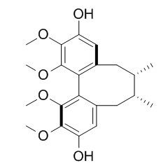
Gomisin J
Gomisin J is a good substrate of cytochrome P450 3A4(CYP3A4),
it has vasodilatory, anti-inflammatory, anti-diabetes, anti-oxidant, and anti-cancer effects, it also has preventive effects on angiotensin II-induced hypertension via an increased nitric oxide bioavailability. Gomisin J has potential benefits in treating nonalcoholic fatty liver disease, it can suppress lipid accumulation by regulating the expression of lipogenic and lipolytic enzymes and inflammatory molecule. Halogenated gomisin J is a potent inhibitor of the cytopathic effects of human immunodeficiency virus type 1 (HIV-1) on MT-4 human T cells (50% effective dose, 0.1 to 0.5 microM).
Inquire / Order:
manager@chemfaces.com
Technical Inquiries:
service@chemfaces.com
Tel:
+86-27-84237783
Fax:
+86-27-84254680
Address:
1 Building, No. 83, CheCheng Rd., Wuhan Economic and Technological Development Zone, Wuhan, Hubei 430056, PRC
Providing storage is as stated on the product vial and the vial is kept tightly sealed, the product can be stored for up to
24 months(2-8C).
Wherever possible, you should prepare and use solutions on the same day. However, if you need to make up stock solutions in advance, we recommend that you store the solution as aliquots in tightly sealed vials at -20C. Generally, these will be useable for up to two weeks. Before use, and prior to opening the vial we recommend that you allow your product to equilibrate to room temperature for at least 1 hour.
Need more advice on solubility, usage and handling? Please email to: service@chemfaces.com
The packaging of the product may have turned upside down during transportation, resulting in the natural compounds adhering to the neck or cap of the vial. take the vial out of its packaging and gently shake to let the compounds fall to the bottom of the vial. for liquid products, centrifuge at 200-500 RPM to gather the liquid at the bottom of the vial. try to avoid loss or contamination during handling.
Chemistry of Plant Materials.2016, 33-46
Anal Chim Acta.2021, 1180:338874.
Nutrients.2017, 10(1)
Plant Cell Physiol.2023, 64(7):716-728.
Food Chemistry: X2023, 101032.
Chulalongkorn University2024, ssrn.4716057.
J Food Sci.2021, 86(9):3810-3823.
Plant Physiol Biochem.2023, 201:107795.
Russian J. Bioorganic Chemistry2024, 50:2897-2903.
Front Chem.2024, 12:1385844.
Related and Featured Products
Vascul Pharmacol. 2012 Sep-Oct;57(2-4):124-30.
Gomisin J from Schisandra chinensis induces vascular relaxation via activation of endothelial nitric oxide synthase.[Pubmed:
22728282]
Gomisin J (GJ) is a lignan contained in Schisandra chinensis (SC) which is a well-known medicinal herb for improvement of cardiovascular symptoms in Korean. Thus, the present study examined the vascular effects of GJ, and also determined the mechanisms involved.
METHODS AND RESULTS:
Exposure of rat thoracic aorta to GJ (1-30μg/ml) resulted in a concentration-dependent vasorelaxation, which was more prominent in the endothelium (ED)-intact aorta. ED-dependent relaxation induced by GJ was markedly attenuated by pretreatment with L-NAME, a nitric oxide synthase (NOS) inhibitor. In the intact endothelial cells of rat thoracic aorta, GJ also enhanced nitric oxide (NO) production. In studies using human coronary artery endothelial cells, GJ enhanced phosphorylation of endothelial NOS (eNOS) at Ser(1177) with increased cytosolic translocation of eNOS, and subsequently increased NO production. These effects of GJ were attenuated not only by calcium chelators including EGTA and BAPTA-AM, but also by LY294002, a PI3K/Akt inhibitor, indicating calcium- and PI3K/Akt-dependent activation of eNOS by GJ. Moreover, the levels of intracellular calcium were increased immediately after GJ administration, but Akt phosphorylation was started to increase at 20min of GJ treatment.
CONCLUSIONS:
Based on these results with the facts that ED-dependent relaxation occurred rapidly after GJ treatment, it was suggested that the ED-dependent vasorelaxant effects of GJ were mediated mainly by calcium-dependent activation of eNOS with subsequent production of endothelial NO.
Antimicrob Agents Chemother. 1995 Sep;39(9):2000-7.
Anti-human immunodeficiency virus (HIV) activities of halogenated gomisin J derivatives, new nonnucleoside inhibitors of HIV type 1 reverse transcriptase.[Pubmed:
8540706]
Halogenated Gomisin J (a derivative of lignan compound), represented by the bromine derivative 1506 [(6R, 7S, S-biar)-4,9-dibromo-3,10-dihydroxy-1,2,11,12-tetramethoxy-6, 7-dimethyl-5,6,7,8- tetrahydrodibenzo[a,c]cyclo-octene], was found to be a potent inhibitor of the cytopathic effects of human immunodeficiency virus type 1 (HIV-1) on MT-4 human T cells (50% effective dose, 0.1 to 0.5 microM).
METHODS AND RESULTS:
Gomisin J derivatives were active in preventing p24 production from acutely HIV-1-infected H9 cells. The selective indices (toxic dose/effective dose) of these compounds were as high as > 300 in some systems. 1506 was active against 3'-azido-3'-deoxythymidine-resistant HIV-1 and acted synergistically with AZT and 2',3'-ddC. 1506 inhibited HIV-1 reverse transcriptase (RT) in vitro but not HIV-1 protease. From the time-of-addition experiment, 1506 was found to inhibit the early phase of the HIV life cycle. A 1506-resistant HIV mutant was selected and shown to possess a mutation within the RT-coding region (at position 188 [Tyr to Leu]). The mutant RT expressed in Escherichia coli was resistant to 1506 in the in vitro RT assay.
Some of the HIV strains resistant to other nonnucleoside HIV-1 RT inhibitors were also resistant to 1506.
CONCLUSIONS:
Comparison of various Gomisin J derivatives with Gomisin J showed that iodine, bromine, and chlorine in the fourth and ninth positions increased RT inhibitory activity as well as cytoprotective activity.
Zhongguo Yao Li Xue Bao. 1996 Nov;17(6):538-41.
Anti-lipid peroxidation of gomisin J on liver mitochondria and cultured myocardial cells.[Pubmed:
9863151]
To study the influences of Gomisin J on lipid peroxidation and calcium paradox.
METHODS AND RESULTS:
Using two in vitro models of rat liver mitochondria membrane lipid peroxidation (LPO) and cultured myocardial cells.
Gomisin J inhibited Fe2+/ascorbic acid and ADP/NADPH-induced LPO with IC50 (95% confidence limits) 5.5 (4.5-6.7) and 4.7 (2.8-7.8) mumol.L-1, respectively, when cultured myocardial cells preincubated with Ca(2+)-free medium for 2 min were incubated with normal medium containing Ca2+, a marked increase of malondialdehyde (MDA) formation occurred and Gomisin J 10 mumol.L-1 protected myocardial cells through decreasing MDA formation.
CONCLUSIONS:
Gomisin J inhibits LPO in rat liver mitochondria and protects cultured myocardial cells from being injured by calcium paradox.
Nat. Prod. Sci., 2006, 12(3):134-7.
Gomisin J with protective effect against t-BHP-induced oxidative damage in HT22 cells from Schizandra chinensis.[Reference:
WebLink]
Four lignan compounds including Gomisin J (1), schizandrin (2), gomisin A (3), and angeloyl gomisin H (4) have been isolated from the MeOH extract of Schizandra chinensis fruits.
METHODS AND RESULTS:
The evaluation for protective effect of compounds 1-4 against tert-butyl hydroperoxide (t-BHP)-induced cytotoxicity in hippocampal HT22 cell line was conducted. Compound 1 showed significant protective effect with an EC50 value of 43.3 ± 2.3 μM, whereas compounds 2-4 were inactive. Trolox, one of the well-known antioxidant, used as a positive control, and also showed protective effect with an EC50 value of 213.8 ± 8.4 μM.
CONCLUSIONS:
These results suggest that compound 1 may possess the neuroprotective activity against oxidant-induced cellular injuries.
Hypertens Res. 2015 Mar;38(3):169-77.
Preventive effect of gomisin J from Schisandra chinensis on angiotensin II-induced hypertension via an increased nitric oxide bioavailability.[Pubmed:
25427681]
Gomisin J (GJ) is a small molecular weight lignan found in Schisandra chinensis and has been demonstrated to have vasodilatory activity.
METHODS AND RESULTS:
In this study, the authors investigated the effect of GJ on blood pressure (BP) in angiotensin II (Ang II)-induced hypertensive mice. In addition, we determined the relative potencies of gomisin A (GA) and GJ with respect to vasodilatory activity and antihypertensive effects. C57/BL6 mice infused s.c. with Ang II (2 μg kg(-1) min(-1) for 2 weeks) showed an increase in BP and a decrease in plasma nitric oxide (NO) metabolites. In the thoracic aortas of Ang II-induced hypertensive mice, a decrease in vascular NO was accompanied by an increase in reactive oxygen species (ROS) production. Furthermore, these alterations in BP, plasma concentrations of NO metabolites and in the vascular productions of NO and ROS in Ang II-treated mice were reversed by the co-administration of GJ (1 and 3 μg kg(-1) min(-1)). In in vitro studies, Ang II decreased the cellular concentration of NO, which was accompanied by a reduction in phosphorylated endothelial nitric oxide synthase (eNOS) and an increase in ROS production. These eNOS phosphorylation and ROS production changes in Ang II-treated cells were also reversed by GJ pretreatment (0-3 μg ml(-1)). Interestingly, the vasodilatory and antihypertensive effects of GJ were more prominent than those of GA.
CONCLUSIONS:
Collectively, an increase in BP in mice treated with Ang II was markedly attenuated by GJ, which was attributed to the preservations of vascular NO bioavailability and eNOS function, and to the inhibition of ROS production in Ang II-induced hypertensive mice.
J Agric Food Chem. 2015 Nov 11;63(44):9729-39.
Gomisin J Inhibits Oleic Acid-Induced Hepatic Lipogenesis by Activation of the AMPK-Dependent Pathway and Inhibition of the Hepatokine Fetuin-A in HepG2 Cells.[Pubmed:
26455261]
The aim of our study is to investigate the molecular mechanism of Gomisin J from Schisandra chinensis on the oleic acid (OA)-induced lipid accumulation in HepG2 cells.
METHODS AND RESULTS:
Gomisin J attenuated lipid accumulation in OA-induced HepG2 cells. It also suppressed the expression of lipogenic enzymes and inflammatory mediators and increased the expression of lipolytic enzymes in OA-induced HepG2 cells. Furthermore, the use of specific inhibitors and fetuin-A siRNA and liver kinase B1 (LKB1) siRNA transfected cells demonstrated that Gomisin J regulated lipogenesis and lipolysis via inhibition of fetuin-A and activation of an AMP-activated protein kinase (AMPK)-dependent pathway in HepG2 cells.Our results showed that Gomisin J suppressed lipid accumulation by regulating the expression of lipogenic and lipolytic enzymes and inflammatory molecules through activation of AMPK, LKB1, and Ca(2+)/calmodulin-dependent protein kinase II and inhibition of fetuin-A in HepG2 cells.
CONCLUSIONS:
This suggested that Gomisin J has potential benefits in treating nonalcoholic fatty liver disease.
Lat. Am. J. Pharm., 2015, 34(2):416-8.
Molecular docking to understand the interaction between anti-tumor drug candidate gomisin J and cytochrome P450 3A4.[Reference:
WebLink]
Protein preparation wizard in the Schrödinger suite of programs was employed to process the structure, and the missing residues and hydrogen atoms were added and assigned.
METHODS AND RESULTS:
Chemdraw software was employed to draw the two-dimensional structure of Gomisin J, and standard bond lengths and angles were given. The ligand of CYP3A4 ketoconazole was firstly extracted, and then the structure of Gomisin J was tried to dock into the active cavity of CYP3A4. The strong hydrogen bonds formed between this compound and the amino acids Ile301 and Ala305 located in the active cavity. To predict the interaction between Gomisin J and the strongest inhibitor of CYP3A4, both Gomisin J and ketoconazole were docked into the activity cavity, and ketoconazole exhibited closer distance with the active site than Gomisin J, indicating ketoconazole can inhibit the metabolism of Gomisin J.
CONCLUSIONS:
In conclusion, the molecular docking indicated that Gomisin J was a good substrate of CYP3A4, and drug-drug interaction between Gomisin J and the inhibitors of CYP3A4 should be given much attention.
Lat. Am. J. Pharm., 2015, 34(4):827-30.
Species-dependent drug-drug interaction between irinotecan and anti-diabetes drug Gomisin J.[Reference:
WebLink]
METHODS AND RESULTS:
Species difference for Gomisin J-irinotecan interaction was determined through determining the inhibition of Gomisin J towards the glucuronidation of the active substance of irinotecan SN-38 in liver microsomes obtained from different species, including human (HLMs), mice (MLMs), and rat (RLMs). Gomisin J (100 uM) was firstly used to screen the inhibition capability of Gomisin J towards the glucuronidation reaction of SN-38 in HLMs, MLMs, and RLMs incubation system. In HLMs and MLMs incubation system, Gomisin J exhibited strong inhibition towards the glucuronidation of SN-38. However, in RLMs incubation system, the inhibition capability is weaker than that in HLMs and MLMs incubation systems. Furthermore, the inhibition kinetic type and parameter were determined.
CONCLUSIONS:
Gomisin J competitively inhibited the glucuronidation of SN-38, and noncompetitively inhibited the glucuronidation of SN-38.
Biosci Biotechnol Biochem. 2010;74(2):285-91.
Anti-inflammatory effects of gomisin N, gomisin J, and schisandrin C isolated from the fruit of Schisandra chinensis.[Pubmed:
20139628]
Schiandra chinensis is a well-known Chinese traditional medicine for the treatment of hepatic disease.
METHODS AND RESULTS:
In this study, we investigated whether the nine major compounds of Schiandra chinensis could be applied to suppress lipopolysaccharide (LPS)-induced inflammatory responses in murine macrophages (Raw 264.7 cells). Among the nine lignans, three, Gomisin J, gomisin N, and schisandrin C, were found to reduce nitric oxide (NO) production from LPS-stimulated Raw 264.7 cells. These three lignans showed low cytotoxic effects in Raw 264.7 cells. Pre-treatment of Raw 264.7 cells with Gomisin J, gomisin N, or schisandrin C reduced the expression of mRNA and the secretion of pro-inflammatory cytokines.
CONCLUSIONS:
These inhibitory effects were found to be caused by blockage of p38 mitogen-activated protein kinase (MAPK), extracellular signal-regulated kinases 1 and 2 (ERK 1/2), and c-Jun N-terminal kinase (JNK) phosphorylation.


