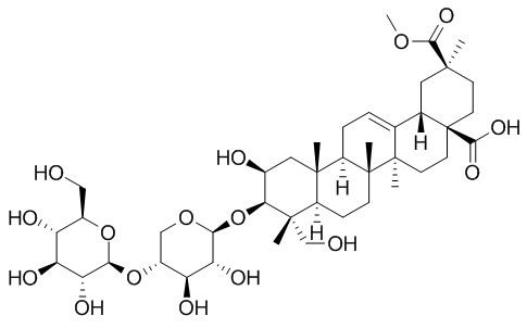
Esculentoside A
Esculentoside A has anti-inflammatory activity , can suppress inflammatory responses in LPS-induced ALI through inhibition of the nuclear factor kappa B and mitogen activated protein kinase signaling pathways. Esculentoside A may be useful for the treatment of autoimmune disease through modulation on T cell-mediated adaptive immunity. Esculentoside A treatment can attenuate CCl4 and GalN/LPS-induced acute liver injury in mice.
Inquire / Order:
manager@chemfaces.com
Technical Inquiries:
service@chemfaces.com
Tel:
+86-27-84237783
Fax:
+86-27-84254680
Address:
1 Building, No. 83, CheCheng Rd., Wuhan Economic and Technological Development Zone, Wuhan, Hubei 430056, PRC
Providing storage is as stated on the product vial and the vial is kept tightly sealed, the product can be stored for up to
24 months(2-8C).
Wherever possible, you should prepare and use solutions on the same day. However, if you need to make up stock solutions in advance, we recommend that you store the solution as aliquots in tightly sealed vials at -20C. Generally, these will be useable for up to two weeks. Before use, and prior to opening the vial we recommend that you allow your product to equilibrate to room temperature for at least 1 hour.
Need more advice on solubility, usage and handling? Please email to: service@chemfaces.com
The packaging of the product may have turned upside down during transportation, resulting in the natural compounds adhering to the neck or cap of the vial. take the vial out of its packaging and gently shake to let the compounds fall to the bottom of the vial. for liquid products, centrifuge at 200-500 RPM to gather the liquid at the bottom of the vial. try to avoid loss or contamination during handling.
Analytical Letters.2020, doi 10.1008
Nutrients.2020, 12(5):1242.
J Sep Sci.2018, 41(7):1682-1690
Applied Physics B2021, 127(92).
VNU J of Science: Med.&Pharm. Sci.2023, 39(1):20-29.
Int J Mol Sci.2018, 19(2)
Pharmacological Reports2020, 1-9
Biomed Pharmacother.2023, 162:114617.
J Pharm Biomed Anal.2024, 251:116444.
Food Analytical Methods2020, 1-10
Related and Featured Products
Bioorg Med Chem Lett. 2007 Dec 1;17(23):6430-3.
Synthesis, in vitro inhibitory activity towards COX-2 and haemolytic activity of derivatives of esculentoside A.[Pubmed:
17950600]
Esculentoside A (EsA) has been reported to possess anti-inflammatory activity and selective inhibitory activity towards cyclooxygenase-2. A series of derivatives of EsA were synthesized by converting the C-28 carboxylic acid group into amides.
METHODS AND RESULTS:
The haemolytic activity and inhibitory activity towards cyclooxygenase-2 were evaluated in vitro. The SAR study of the derivatives was conducted and showed that introducing aromatic ring to EsA greatly enhanced its biological activity.
CONCLUSIONS:
Compound 23 showed higher inhibitory activity than Celecoxib and EsA, but lower haemolytic toxicity than EsA.
J Surg Res. 2013 Nov;185(1):364-72.
Protective effect of esculentoside A on lipopolysaccharide-induced acute lung injury in mice.[Pubmed:
23764313]
Esculentoside A (EsA) is a saponin isolated from the Chinese herb Phytolacca esculenta. In our study, we sought to investigate the protective effects of Esculentoside A on lipopolysaccharide (LPS)-induced acute lung injury (ALI) in mice.
METHODS AND RESULTS:
To determine the effects of Esculentoside A on the reduction of histopathologic changes in mice with ALI, inflammatory cell count in bronchoalveolar lavage fluid (BALF) and lung wet-to-dry weight ratio were measured in LPS-challenged mice, and lung histopathologic changes observed via paraffin section were assessed. Next, cytokine production induced by LPS in BALF was measured by enzyme-linked immunosorbent assay. To further study the mechanism of Esculentoside A protective effects on ALI, IκBa, p38, and extracellular signal receptor-activated kinase pathways were investigated in lung tissue of mice with ALI. In the present investigation, Esculentoside A showed marked effects by reducing inflammatory infiltration, thickening of the alveolar wall, and pulmonary congestion. Levels of tumor necrosis factor α and interleukin 6 elevated by LPS were significantly decreased in BALF in Esculentoside A-pretreated ALI model. Furthermore, Esculentoside A significantly suppressed phosphorylation of IκBa, p38, and extracellular signal receptor-activated kinase.
CONCLUSIONS:
Taken together, our results suggest that Esculentoside A suppressed inflammatory responses in LPS-induced ALI through inhibition of the nuclear factor kappa B and mitogen activated protein kinase signaling pathways. Esculentoside A may be a promising potential preventive agent for ALI treatment.
Arch Med Sci. 2013 Apr 20;9(2):354-60.
The effect of esculentoside A on lupus nephritis-prone BXSB mice.[Pubmed:
23671449]
Esculentoside A was reported to have the effect of modulating immune response, cell proliferation and apoptosis as well as anti-inflammatory effects in acute and chronic experimental models. However, the effects of Esculentoside A on LN remain poorly understood. To investigate the roles of Esculentoside A in LN, the effects of Esculentoside A were tested on BXSB mice, a SLE model, in which male SB/Le mice and female C57BL/6 mice were hybridized through recombinant inbred species.
METHODS AND RESULTS:
Twenty four BXSB mice were divided into three groups. After 4 weeks, blood samples, urine samples and kidney tissues were collected. Measurement of cytokine levels was carried out using sandwich Esculentoside A reagent kits. Apoptotic scores were obtained with a TUNEL assay. PCNA and Caspase-3 mRNA was detected using the In Situ Hybridization Detection Kit. The results demonstrated that compared with the control group, Esculentoside A administration markedly controlled urine protein excretion, improved renal function, alleviated kidney damage and promoted the apoptosis of glomerular intrinsic cells and renal tubular epithelial cells in animals of the treated group (p < 0.05). Meanwhile, Esculentoside A reduced the serum IL-6 and TNF-α levels (p < 0.05), inhibited the expression of PCNA and promoted the expression of caspase-3, Fas and FasL in animals of the treated group (p < 0.05). The effects of Esculentoside A on BXSB mice were similar to dexamethasone.
CONCLUSIONS:
All these findings indicated that Esculentoside A might play significant roles in the treatment of BXSB mice through modulation of inflammatory cytokines, inhibition of renal cell proliferation and induction of apoptosis. The special targets of Esculentoside A in lupus nephritis are worth further exploration.
Pharmacology. 1998 Apr;56(4):187-95.
Esculentoside A inhibits tumor necrosis factor, interleukin-1, and interleukin-6 production induced by lipopolysaccharide in mice.[Pubmed:
9566020]
Esculentoside A, a kind of saponin isolated from the root of the Chinese herb Phytolaca esculenta, is reported to possess potent anti-inflammatory effects in acute and chronic experimental models.
METHODS AND RESULTS:
In the present study, we investigated the effects of Esculentoside A on the production of tumor necrosis factor (TNF), interleukin-1 (IL-1) and interleukin-6 (IL-6) induced by lipopolysaccharide (LPS) in mice. In vitro experiments demonstrated that Esculentoside A (0.1-10 mumol/l) significantly reduced the release of TNF from the peritoneal macrophages derived from mice pretreated with thioglycolate. IL-1 and IL-6 secretion was also obviously inhibited in a concentration-dependent manner by Esculentoside A from 0.01 to 10 mumol/l. In vivo experiments demonstrated that detectable TNF was observed 0.25 h after injection, was maximal at 0.5 h, and returned to baseline at 4 h. Maximal production of IL-1 and IL-6 were observed to be 1 and 2 h, respectively, after injection of LPS. Pretreatment of mice with 5, 10, or 20 mg/kg Esculentoside A once a day for 7 consecutive days dose-dependently decreased the TNF, IL-1 and IL-6 levels in the sera of mice following LPS challenge. TNF, IL-1, and IL-6 are important cytokines involved in the pathogenesis of inflammatory lesions.
CONCLUSIONS:
Inhibition of inflammatory cytokine production may contribute to the anti-inflammatory effects of Esculentoside A.
PLoS One. 2014 Nov 18;9(11):e113107.
The protective effect of esculentoside a on experimental acute liver injury in mice.[Pubmed:
25405982]
Inflammatory response and oxidative stress are considered to play an important role in the development of acute liver injury induced by carbon tetrachloride (CCl4) and galactosamine (GalN)/lipopolysaccharides (LPS). Esculentoside A (EsA), isolated from the Chinese herb phytolacca esculenta, has the effect of modulating immune response, cell proliferation and apoptosis as well as anti-inflammatory effects.
METHODS AND RESULTS:
The present study is to evaluate the protective effect of EsA on CCl4 and GalN/LPS-induced acute liver injury. In vitro, CCK-8 assays showed that EsA had no cytotoxicity, while it significantly reduced levels of TNF-α and cell death rate challenged by CCl4. Moreover, EsA treatment up-regulated PPAR-γ expression of LO2 cells and reduced levels of reactive oxygen species (ROS) challenged by CCl4. In vivo, EsA prevented mice from CCl4-induced liver histopathological damage. In addition, levels of AST and ALT were significantly decreased by EsA treatment. Furthermore, the mice treated with EsA had a lower level of TNF-α, Interleukin (IL)-1β and IL-6 in mRNA expression. EsA prevented MDA release and increased GSH-Px activity in liver tissues. Immunohistochemical staining showed that over-expression of F4/80 and CD11b were markedly inhibited by EsA. The western bolt results showed that EsA significantly inhibited CCl4-induced phosphonated IkBalpha (P-IκB) and ERK. Furthermore, EsA treatment also alleviated GalN/LPS-induced acute liver injury on liver enzyme and histopathological damage. Unfortunately, our results exhibited that EsA had no effects on CCl4-induced hepatocyte apoptosis which were showed by TUNEL staining and Bax, Caspase-3 and cleaved Caspase-3 expression.
CONCLUSIONS:
Our results proved that EsA treatment attenuated CCl4 and GalN/LPS-induced acute liver injury in mice and its protective effects might be involved in inhibiting inflammatory response and oxidative stress, but not apoptosis with its underlying mechanism associated with PPAR-γ, NF-κB and ERK signal pathways.
Int Immunopharmacol. 2010 Jan;10(1):65-71.
Effects of esculentoside A on autoimmune syndrome induced by Campylobacterjejuni in mice and its modulation on T-lymphocyte proliferation and apoptosis.[Pubmed:
19818416]
Esculentoside A (EsA), a saponin isolated from the root of Phytolacca esculenta, has been reported to exert anti-inflammatory effects in several animal models of acute and chronic inflammation by inhibiting the production and activity of pro-inflammatory cytokines in macrophages and epithelial cells. However, little is known about its modulation on T cells.
METHODS AND RESULTS:
In the present study, we further investigated its potential in treatment of autoimmune disease and its modulation on T cells, using an experimental autoimmune model established through immunizing mice with Campylobacterjejuni strain CJ-S(131) in Freund's complete adjuvant. Our results demonstrated that EsA administration markedly alleviated the inflammatory injury in liver and kidney of model mice, decreased the anti-CD3/CD28-stimulated proliferation of splenocytes and lymph node cells, and reduced the percentage of CD3+, CD4+, and CD8+ lymphocytes in peripheral blood. Furthermore, we demonstrated that EsA induced apoptosis in ConA-activated thymocytes but not in non-activated thymocytes. Gene expression analysis revealed that EsA up-regulated the expression of a group of pro-apoptotic genes more profoundly in Con A-activated thymocytes than in non-activated thymocytes. EsA-affected pro-apoptotic genes included those involved in Fas induction, p53 activation, redox metabolism, calcium- and glucocorticoid-induced apoptosis signals, suggesting that EsA may modulate multiple apoptotic signal pathways in activated T cells.
CONCLUSIONS:
Taken together, our findings suggest that EsA may be useful for the treatment of autoimmune disease through modulation on T cell-mediated adaptive immunity.


