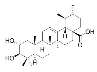
Corosolic acid
Corosolic acid has antitumor, anti-inflammatory and hypoglycemic activities, it can ameliorate hypertension, abnormal lipid metabolism, and oxidative stress as well as the inflammatory state in SHR-cp rats; it can improve glucose metabolism by reducing insulin resistance, it inhibits the enzymatic activities of several diabetes-related non-receptor protein tyrosine phosphatases (PTPs) in vitro, such as PTP1B, T-cell-PTP, src homology phosphatase-1 and src homology phosphatase-2. Corosolic acid can suppress the M2 polarization of macrophages and tumor cell proliferation by inhibiting both STAT3 and NF-κB activation, it also can enhance the antitumor effects of adriamycin and cisplatin in in vitro.
Inquire / Order:
manager@chemfaces.com
Technical Inquiries:
service@chemfaces.com
Tel:
+86-27-84237783
Fax:
+86-27-84254680
Address:
1 Building, No. 83, CheCheng Rd., Wuhan Economic and Technological Development Zone, Wuhan, Hubei 430056, PRC
Providing storage is as stated on the product vial and the vial is kept tightly sealed, the product can be stored for up to
24 months(2-8C).
Wherever possible, you should prepare and use solutions on the same day. However, if you need to make up stock solutions in advance, we recommend that you store the solution as aliquots in tightly sealed vials at -20C. Generally, these will be useable for up to two weeks. Before use, and prior to opening the vial we recommend that you allow your product to equilibrate to room temperature for at least 1 hour.
Need more advice on solubility, usage and handling? Please email to: service@chemfaces.com
The packaging of the product may have turned upside down during transportation, resulting in the natural compounds adhering to the neck or cap of the vial. take the vial out of its packaging and gently shake to let the compounds fall to the bottom of the vial. for liquid products, centrifuge at 200-500 RPM to gather the liquid at the bottom of the vial. try to avoid loss or contamination during handling.
Molecules.2018, 23(12):E3103
Phytomedicine.2021, 93:153796.
Foods.2023, 12(7):1355.
Foods.2023, 12(6):1130.
Virulence.2018, 9(1):588-603
BMC Biotechnol.2024, 24(1):94.
Naunyn Schmiedebergs Arch Pharmacol.2024, 03148-x.
bioRxiv - Molecular Biology2023, 535548.
Biol Pharm Bull.2021, 44(12):1891-1893.
Talanta.2022, 249:123645.
Related and Featured Products
Nat Prod Res. 2014;28(21):1879-86.
Microbial transformation of the anti-diabetic agent corosolic acid.[Pubmed:
25190540]
METHODS AND RESULTS:
Biotransformation of Corosolic acid (1) by Cochliobolus lunatus and Streptomyces asparaginoviolaceus afforded four metabolites, which were identified by using (1)H NMR, (13)C NMR, DEPT, HSQC, HMBC and NOESY spectral data. Biotransformation of Corosolic acid by C. lunatus R.R. Nelson & Haasis CGMCC 3.4381 produced three metabolites: 2α,3β,21β-trihydroxyurs-12-en-28-oic acid (2), 2α,3β,7β,21β-tetrahydroxy-urs-12-en-28-oic acid (3) and 2α,3β-dihydroxy-21-oxours-12-en-28-oic acid (4). Incubation of Corosolic acid with growing cultures of S. asparaginoviolaceus CGMCC 4.0175 afforded metabolite 2α,3β,30-trihydroxyurs-12-en-28-oic acid (5). All the metabolites were reported for the first time.
CONCLUSIONS:
The substrate and four metabolites, along with four products obtained previously, were evaluated for their inhibitory effects on α-glucosidase; all the triterpenes tested showed potent inhibitory effects.
Phytother Res. 2015 May;29(5):714-23.
Corosolic Acid Exhibits Anti-angiogenic and Anti-lymphangiogenic Effects on In Vitro Endothelial Cells and on an In Vivo CT-26 Colon Carcinoma Animal Model.[Pubmed:
25644809]
We describe the anti-angiogenic and anti-lymphangiogenic effects of Corosolic acid, a pentacyclic triterpenoid isolated from Cornus kousa Burg.
METHODS AND RESULTS:
A mouse colon carcinoma CT-26 animal model was employed to determine the in vivo anti-angiogenic and anti-lymphangiogenic effects of Corosolic acid. Corosolic acid induced apoptosis in CT-26 cells, mediated by the activation of caspase-3. In addition, it reduced the final tumor volume and the blood and lymphatic vessel densities of tumors, indicating that it suppresses in vivo angiogenesis and lymphangiogenesis. Corosolic acid inhibited the proliferation and tube formation of human umbilical vein endothelial cells and human dermal lymphatic microvascular endothelial cells. In addition, Corosolic acid decreased the proliferation and migration of human umbilical vein endothelial cells stimulated by angiopoietin-1.
CONCLUSIONS:
Pretreatment with Corosolic acid decreased the phosphorylation of focal adhesion kinase (FAK) and ERK1/2, suggesting that Corosolic acid contains anti-angiogenic activity that can suppress FAK signaling induced by angiopoietin-1.
Food Chem Toxicol. 2014 May;67:87-95.
Ursolic acid and its natural derivative corosolic acid suppress the proliferation of APC-mutated colon cancer cells through promotion of β-catenin degradation.[Pubmed:
24566423]
Ursolic acid (UA) and Corosolic acid (CA), naturally occurring pentacyclic triterpene acids, exhibit antiproliferative activities against various cancer cells, but a clear chemopreventive mechanism of these triterpenoids in colon cancer cells remains to be answered.
METHODS AND RESULTS:
Here we used a cell-based reporter system for detection of β-catenin response transcription (CRT) to identify UA as an antagonist of the Wnt/β-catenin pathway. UA promoted the degradation of intracellular β-catenin that was accompanied by its N-terminal phosphorylation at Ser33/37/Thr41 residues, marking it for proteasomal degradation. Consistently, UA down-regulated the intracellular β-catenin level in colon cancer cells with inactivating mutations of adenomatous polyposis coli (APC). In addition, UA repressed the expression of β-catenin/T-cell factor (TCF)-dependent genes, thereby inhibiting cell proliferation in colon cancer cells. The functional group analysis revealed that the major structural requirements for UA-mediated β-catenin degradation are a carboxyl group at position 17 and a methyl group at position 19. Notably, CA (2α-hydroxyursolic acid) was also found to decrease the level of intracellular β-catenin and to suppress the growth of APC-mutated colon cancer cells.
CONCLUSIONS:
Our findings suggest that UA and CA exert their anticancer activities against colon cancer cells by promoting the N-terminal phosphorylation and subsequent proteasomal degradation of β-catenin.
PLoS One. 2015 May 15;10(5):e0126725.
Corosolic Acid Inhibits Hepatocellular Carcinoma Cell Migration by Targeting the VEGFR2/Src/FAK Pathway.[Pubmed:
25978354 ]
Inhibition of VEGFR2 activity has been proposed as an important strategy for the clinical treatment of hepatocellular carcinoma (HCC).
METHODS AND RESULTS:
In this study, we identified Corosolic acid (CA), which exists in the root of Actinidia chinensis, as having a significant anti-cancer effect on HCC cells. We found that CA inhibits VEGFR2 kinase activity by directly interacting with the ATP binding pocket. CA down-regulates the VEGFR2/Src/FAK/cdc42 axis, subsequently decreasing F-actin formation and migratory activity in vitro. In an in vivo model, CA exhibited an effective dose (5 mg/kg/day) on tumor growth. We further demonstrate that CA has a synergistic effect with sorafenib within a wide range of concentrations.
CONCLUSIONS:
In conclusion, this research elucidates the effects and molecular mechanism for CA on HCC cells and suggests that CA could be a therapeutic or adjuvant strategy for patients with aggressive HCC.
Int J Mol Med. 2014 Apr;33(4):943-9.
Corosolic acid induces apoptotic cell death in HCT116 human colon cancer cells through a caspase-dependent pathway.[Pubmed:
24481288]
Corosolic acid (CA), a pentacyclic triterpene isolated from Lagerstroemia speciosa L. (also known as Banaba), has been shown to exhibit anticancer properties in various cancer cell lines. However, the anticancer activity of CA on human colorectal cancer cells and the underlying mechanisms remain to be elucidated.
METHODS AND RESULTS:
In this study, we investigated the effects of CA on cell viability and apoptosis in HCT116 human colon cancer cells. CA dose-dependently inhibited the viability of HCT116 cells. The typical hallmarks of apoptosis, such as chromatin condensation, a sub-G1 peak and phosphatidylserine externalization were detected by Hoechst 33342 staining, flow cytometry and Annexin V staining following treatment with CA. Western blot analysis revealed that CA induced a decrease in the levels of procaspase-8, -9 and -3 and the cleavage of poly(ADP-ribose) polymerase (PARP). The apoptotic cell death induced by CA was accompanied by the activation of caspase-8, -9 and -3, which was completely abrogated by the pan-caspase inhibitor, z-VAD‑FMK. Furthermore, CA upregulated the levels of pro-apoptotic proteins, such as Bax, Fas and FasL and downregulated the levels of anti-apoptotic proteins, such as Bcl-2 and survivin.
CONCLUSIONS:
Taken together, our data provide insight into the molecular mechanisms of CA-induced apoptosis in colorectal cancer (CRC), rendering this compound a potential anticancer agent for the treatment of CRC.


