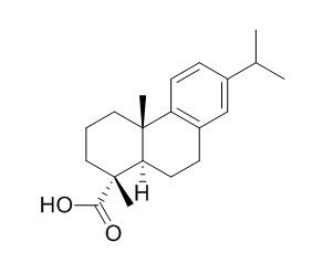
Dehydroabietic acid
Dehydroabietic acid , a major poison to fishes in pulp and paper mill effluents, which
could be useful in improving the diabetic wound healing, it can reverse several cell responses stimulated by TNF-α, including the activation of FOXO1 and the TGF-β1/Smad3 signaling pathway. Dehydroabietic acid derivatives displays antisecretory and antipepsin effect, have gastroprotective activity in the HCl/EtOH-induced gastric lesions in mice as well as for cytotoxicity in human lung fibroblasts (MRC-5) and human epithelial gastric (AGS) cells.
Inquire / Order:
manager@chemfaces.com
Technical Inquiries:
service@chemfaces.com
Tel:
+86-27-84237783
Fax:
+86-27-84254680
Address:
1 Building, No. 83, CheCheng Rd., Wuhan Economic and Technological Development Zone, Wuhan, Hubei 430056, PRC
Providing storage is as stated on the product vial and the vial is kept tightly sealed, the product can be stored for up to
24 months(2-8C).
Wherever possible, you should prepare and use solutions on the same day. However, if you need to make up stock solutions in advance, we recommend that you store the solution as aliquots in tightly sealed vials at -20C. Generally, these will be useable for up to two weeks. Before use, and prior to opening the vial we recommend that you allow your product to equilibrate to room temperature for at least 1 hour.
Need more advice on solubility, usage and handling? Please email to: service@chemfaces.com
The packaging of the product may have turned upside down during transportation, resulting in the natural compounds adhering to the neck or cap of the vial. take the vial out of its packaging and gently shake to let the compounds fall to the bottom of the vial. for liquid products, centrifuge at 200-500 RPM to gather the liquid at the bottom of the vial. try to avoid loss or contamination during handling.
Eur J Pharmacol.2024, 975:176644.
J Nat Prod.2022, 85(5):1351-1362.
Pharmaceuticals (Basel).2024, 17(6):727.
Chemistry of Natural Compounds2018, 204-206
J Biol Chem.2014, 289(3):1723-31
Food Bioscience2023, 56:103311.
Int J Mol Sci.2021, 22(16):8641.
Phytomedicine.2022, 100:154085.
Nutrients.2020, 12(5):1242.
Food Chem.2018, 252:207-214
Related and Featured Products
Int J Clin Exp Pathol. 2014 Dec 1;7(12):8616-26.
Dehydroabietic acid reverses TNF-α-induced the activation of FOXO1 and suppression of TGF-β1/Smad signaling in human adult dermal fibroblasts.[Pubmed:
25674226]
Wound healing impairment is a well-documented phenomenon in clinical and experimental diabetes, and in diabetic wound healing impaired fibroblast has been linked to increased levels of tumor necrosis factor-α (TNF-α).
METHODS AND RESULTS:
A number of signaling pathways including TNF-α/forkhead box O1 (FOXO1) and transforming growth factor-β1 (TGF-β1)/Smads in fibroblasts appear to play a cardinal role in diabetic wound healing. Dehydroabietic acid (DAA) is obtained from Commiphora oppbalsamum and inhibits the production of TNF-α in macrophages and adipocytes, decreases the level of TNF-α in obese diabetic KK-Ay mice, but its effect on diabetic wound healing is unknown. This study was to investigate the effect of DAA on TNF-α-stimulated human adult dermal fibroblasts. On the one hand, TNF-α significantly decreased the fibroblast proliferation and the expression of PCNA, Ki67 and cyclin D1, increased the fibroblast apoptosis, caspase-8/3 activity, expressions of cleaved caspase-8 and caspase-3, decreased the Bcl-2/Bax ratio and increased activation of the pro-apoptotic transcription factor FOXO1. All the above-mentioned cell responses were remarkably reversed by DAA. On the other hand, TNF-α also inhibited TGF-β1-induced the Smad3 signaling pathway what is closely related to the fibroblast migration and the differentiation of myofibroblasts. However, DAA significantly promoted the migration and increased the expression of α-smooth muscle actin and fibronectin under the stimulus of a combination of TNF-α and TGF-β1.
CONCLUSIONS:
In conclusion, DAA could reverse several cell responses stimulated by TNF-α, including the activation of FOXO1 and the TGF-β1/Smad3 signaling pathway. These results suggested that DAA could be useful in improving the diabetic wound healing.
Pharmacol Res. 2005 Nov;52(5):429-37.
Gastroprotective and cytotoxic effect of dehydroabietic acid derivatives.[Pubmed:
16125407 ]
Dehydroabietic acid derivatives have been reported to display antisecretory and antipepsin effect in animal models. Some 19 Dehydroabietic acid diterpenes were prepared and assessed for gastroprotective activity in the HCl/EtOH-induced gastric lesions in mice as well as for cytotoxicity in human lung fibroblasts (MRC-5) and human epithelial gastric (AGS) cells.
METHODS AND RESULTS:
At a single oral dose of 100 mg kg(-1), highest gastroprotective effect was provided by dehydroabietanol, its corresponding aldehyde, Dehydroabietic acid (DHA) and its methyl ester, N-(m-nitrophenyl)-, N-(o-chlorophenyl)- and N-(p-iodophenyl)abieta-8,11,13-trien-18-amide (compounds 12-14), N-2-aminothiazolyl- and N-benzylabieta-8,11,13-trien-18-amide (compounds 18-19) being as active as lansoprazole at 20 mg kg(-1) and reducing the lesion index by at least 75%. In the compound series including the alcohol, ester, aldehyde, acid and methyl ester at C-18 (compounds 1-9), highest activity was related with the presence of an alcohol, aldehyde, acid or methyl ester at C-18, the activity being strongly reduced after esterification. The cytotoxicity of the compounds 1-9 towards AGS cells and fibroblasts was higher than the values for the amides 10-19. In the compounds 10-19, the best gastroprotective effect was observed for the aromatic amides 12-14 (75-85% inhibition of gastric lesions) bearing a nitro or halogen function in the N-benzoyl moiety. Lowest cytotoxicity was found for the amides, with IC(50) values >1000 microM for fibroblasts and from 200 up to >1000 microM for AGS cells, respectively.
CONCLUSIONS:
The N-2-aminothiazolyl- and N-benzylamide derivatives were also very active as gastroprotectors with higher cytotoxicity against AGS cells.
Biomed Res Int. 2014;2014:682197.
Dehydroabietic acid derivative QC2 induces oncosis in hepatocellular carcinoma cells.[Pubmed:
25110686]
Rosin, the traditional Chinese medicine, is reported to be able to inhibit skin cancer cell lines. In this report, we investigate the inhibitory effect against HCC cells of QC2, the derivative of rosin's main components Dehydroabietic acid.
METHODS AND RESULTS:
MTT assay was used to determine the cytotoxicity of QC2. Morphological changes were observed by time-lapse microscopy and transmission electron microscopy and the cytoskeleton changes were observed by laser-scanning confocal microscopy. Cytomembrane integrity and organelles damage were confirmed by detection of the reactive oxygen (ROS), lactate dehydrogenase (LDH), and mitochondrial membrane potential (Δψm). The underlying mechanism was manifested by Western blotting. The oncotic cell death was further confirmed by detection of oncosis related protein calpain. RESULTS: Swelling cell type and destroyed cytoskeleton were observed in QC2-treated HCC cells. Organelle damage was visualized by transmission electron microscopy. The detection of ROS accumulation, increased LDH release, and decreased ATP and Δψm confirmed the cell death. The oncotic related protein calpain was found to increase time-dependently in QC2-treated HCC cells, while its inhibitor PD150606 attenuated the cytotoxicity.
CONCLUSIONS:
Dehydroabietic acid derivative QC2 activated oncosis related protein calpain to induce the damage of cytomembrane and organelles which finally lead to oncosis in HCC cells.
Water Res., 1983, 17(1):81-9.
Toxicological effects of dehydroabietic acid (DHAA) on the trout, Salmo gairdneri Richardson, in fresh water.[Reference:
WebLink]
METHODS AND RESULTS:
Toxicological and physiological effects of Dehydroabietic acid (DHAA), a major poison to fishes in pulp and paper mill effluents, were studied by two experiments with rainbow trout, Salmo gairdneri Richardson: in the first, fish were acutely exposed for 4 days to an average DHAA concentration of 1.2 mg l−1 (Exp. I) and in the second for 30 days to an average of 20 μg DHAA l−1 (Exp. II). Compared to the controls, fish of Exp. I displayed a decreased relative weight of liver, an increased blood haematocrit, and increased haemoglobin as well as plasma protein concentrations. The aspartate aminotransferase activity of heart muscle was significantly elevated, as was also the lactate dehydrogenase (LDH) of white muscle tissue. In the blood plasma, the proportion of muscle type LDH activity was simultaneously increased. UDP-glucuronyl-transferase activities of liver and kidney were strongly decreased. Results suggest an increased and altered use of body energy reserves, decreased plasma volume and impaired liver function. Fish of Exp. II showed an increased relative weight of spleen. In addition, liver and gill LDH shifted towards heart-type.
CONCLUSIONS:
We conclude that 20 μg l−1 is close to the “minimum effective concentration” of DHAA to rainbow trout.


