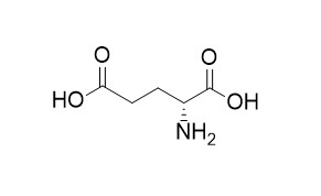
D-Glutamic acid
Poly-D-glutamic acid induces an acute lysosomal thesaurismosis of proximal tubules and a marked proliferation of interstitium in rat kidney.
Inquire / Order:
manager@chemfaces.com
Technical Inquiries:
service@chemfaces.com
Tel:
+86-27-84237783
Fax:
+86-27-84254680
Address:
1 Building, No. 83, CheCheng Rd., Wuhan Economic and Technological Development Zone, Wuhan, Hubei 430056, PRC
Providing storage is as stated on the product vial and the vial is kept tightly sealed, the product can be stored for up to
24 months(2-8C).
Wherever possible, you should prepare and use solutions on the same day. However, if you need to make up stock solutions in advance, we recommend that you store the solution as aliquots in tightly sealed vials at -20C. Generally, these will be useable for up to two weeks. Before use, and prior to opening the vial we recommend that you allow your product to equilibrate to room temperature for at least 1 hour.
Need more advice on solubility, usage and handling? Please email to: service@chemfaces.com
The packaging of the product may have turned upside down during transportation, resulting in the natural compounds adhering to the neck or cap of the vial. take the vial out of its packaging and gently shake to let the compounds fall to the bottom of the vial. for liquid products, centrifuge at 200-500 RPM to gather the liquid at the bottom of the vial. try to avoid loss or contamination during handling.
Int J Mol Sci.2020, 21(19),7070.
Phytochem Anal.2023, pca.3305.
J. Korean Wood Sci. Technol.2022, 50(5):338-352.
Heliyon.2024, 10(11):e32352.
Geroscience.2024, 01207-y.
Molecules.2019, 25(1):E103
Heliyon.2024, 10(7):e28755.
Phytomedicine.2024, 129:155645.
Srinagarind Medical Journal2019, 34(1)
J Pharm Biomed Anal.2021, 196:113931.
Related and Featured Products
Laboratory Investigation, 1996, 74(6):1013-1023.
Poly- D-glutamic acid induced an acute lysosomal thesaurismosis of proximal tubules and a marked proliferation of interstitium in rat kidney.[Reference:
WebLink]
METHODS AND RESULTS:
Renal damage caused by polycationic peptides is well documented, but renal damage caused by polyanionic peptides is not. During our attempts to inhibit the nephrotoxicity of aminoglycoside antibiotics by polyanionic peptides, we discovered that poly-D-Glutamic acid (molecular weight, 20 kd; 250 mg/kg/day subcutaneously for 1 to 4 days) produces an acute thesaurismosis in the proximal tubular cells associated with a marked proliferation of peritubular interstitial cells in rat kidney. Thesaurismotic bodies were easily visualized by light microscopy at the basal pole of proximal tubular cells with the cationic stain Giemsa. By electron microscopy, these bodies appeared membrane-limited, frequently distorted, filled with heterogeneous granular material, accessible to injected peroxidase (a tracer of the endocytic pathway), and generally stainable for the lysosomal enzyme arylsulfatase. Specimens obtained 3 hours after injection of poly-D-Glutamic acid and horseradish peroxidase suggested an impairment of endosome and/or lysosome fission, but not fusion. By histoautoradiographic examination after 3H-thymidine incorporation, global labeling indices of cortical cells were increased 11- to 18-fold in poly-D-Glutamic acid-treated rats as compared with controls, with > 80% of labeled cells localized in the interstitium. Distal tubular and glomerular cells also showed a moderate proliferation, but proximal tubular cells showed no significant necrosis or proliferation. Although tubular thesaurismosis persisted, interstitial cell proliferation resolved within 7 days after cessation of treatment.
CONCLUSIONS:
We suggest that poly-D-Glutamic acid is a convenient tool to induce a rapid and sustained lysosomal storage disorder. It could also help clarify the relationship between insults to tubular cells and proliferation of peritubular cells, two features frequently associated in tubulointerstitial disorders. The mechanism of the thesaurismosis and of the interference with the dynamics of fusion-fission of the endocytic apparatus are addressed in the companion paper.
Journal of Chromatography B, 2011, 879(29):3196-3202.
Simultaneous determination of d-aspartic acid and d-glutamic acid in rat tissues and physiological fluids using a multi-loop two-dimensional HPLC procedure.[Reference:
WebLink]
For a metabolomics study focusing on the analysis of aspartic and glutamic acid enantiomers, a fully automated two-dimensional HPLC system employing a microbore-ODS column and a narrowbore-enantioselective column was developed.
METHODS AND RESULTS:
By using this system, a detailed distribution of d-Asp and d-Glu besides l-Asp and l-Glu in mammals was elucidated. For the total analysis concept, the amino acids were first pre-column derivatized with 4-fluoro-7-nitro-2,1,3-benzoxadiazole (NBD-F) to be sensitively and fluorometrically detected. For the non-stereoselective separation of the analytes in the first dimension a monolithic ODS column (750 mm × 0.53 mm i.d.) was adopted, and a self-packed narrowbore-Pirkle type enantioselective column (Sumichiral OA-2500S, 250 mm × 1.5 mm i.d.) was selected for the second dimension. In the rat plasma, RSD values for intra-day and inter-day precision were less than 6.8%, and the accuracy ranged between 96.1% and 105.8%. The values of LOQ of d-Asp and d-Glu were 5 fmol/injection (0.625 nmol/g tissue).
CONCLUSIONS:
The present method was successfully applied to the simultaneous determination of free aspartic acid and glutamic acid enantiomers in 7 brain areas, 11 peripheral tissues, plasma and urine of Wistar rats. Biologically significant d-Asp values were found in various tissue samples whereas for d-Glu(D-Glutamic acid) the values were very low possibly indicating less significance.
Journal of Bacteriology, 1993, 175(1):111-116.
The Escherichia coli mutant requiring D-glutamic acid is the result of mutations in two distinct genetic loci.[Reference:
WebLink]
D-Glutamic acid is an essential component of bacterial cell wall peptidoglycan in both gram-positive and gram-negative bacteria. Very little is known concerning the genetics and biochemistry of D-glutamate production in most bacteria, including Escherichia coli.
METHODS AND RESULTS:
Evidence is presented in this report for the roles of two distinct genes in E. coli WM335, a strain which is auxotrophic for D-glutamate. The first gene, which restores D-glutamate independence in WM335, was mapped, cloned, and sequenced. This gene, designated dga, is a previously reported open reading frame, located at 89.8 min on the E. coli map. The second gene, gltS, is located at 82 min. gltS encodes a protein that is involved in the transport of D- and L-glutamic acid into E. coli, and the gltS gene of WM335 was found to contain two missense mutations.
CONCLUSIONS:
To construct D-glutamate auxotrophs, it is necessary to transfer sequentially the mutated gltS locus, and then the mutated dga locus into the recipient. The sequences of the mutant forms of both dga and gltS are also presented.


