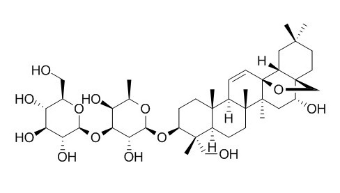
Saikosaponin D
Saikosaponin D, a calcium mobilizing agent (SERCA inhibitor), is also an agonist of the glucocorticoid receptor (GR),which has anti-cancer, anti-inflammatory, and neuroprotective effects. Saikosaponin D protects against acetaminophen-induced hepatotoxicity by inhibiting NF-κB and STAT3 signaling; it shows inhibitory effects on NF-κB activation and thereby on iNOS, COX-2 and pro-inflammatory cytokines.
Inquire / Order:
manager@chemfaces.com
Technical Inquiries:
service@chemfaces.com
Tel:
+86-27-84237783
Fax:
+86-27-84254680
Address:
1 Building, No. 83, CheCheng Rd., Wuhan Economic and Technological Development Zone, Wuhan, Hubei 430056, PRC
Providing storage is as stated on the product vial and the vial is kept tightly sealed, the product can be stored for up to
24 months(2-8C).
Wherever possible, you should prepare and use solutions on the same day. However, if you need to make up stock solutions in advance, we recommend that you store the solution as aliquots in tightly sealed vials at -20C. Generally, these will be useable for up to two weeks. Before use, and prior to opening the vial we recommend that you allow your product to equilibrate to room temperature for at least 1 hour.
Need more advice on solubility, usage and handling? Please email to: service@chemfaces.com
The packaging of the product may have turned upside down during transportation, resulting in the natural compounds adhering to the neck or cap of the vial. take the vial out of its packaging and gently shake to let the compounds fall to the bottom of the vial. for liquid products, centrifuge at 200-500 RPM to gather the liquid at the bottom of the vial. try to avoid loss or contamination during handling.
J Agric Food Chem.2017, 65(13):2670-2676
Appl. Sci.2023, 13(2), 860.
Molecules.2024, 29(23):5632.
Phytomedicine.2017, 24:77-86
Microchemical Journal2023. 191:108938
World J Mens Health.2019, 10.5534
Sains Malaysiana2024, 53(2):397-408.
Food Science and Biotechnology2015, 2205-2212
Plants (Basel).2020, 9(11):1555.
Nat Prod Commun.2014, 9(5):679-82
Related and Featured Products
Eur Rev Med Pharmacol Sci. 2014;18(17):2435-43.
Saikosaponin-d inhibits proliferation of human undifferentiated thyroid carcinoma cells through induction of apoptosis and cell cycle arrest.[Pubmed:
25268087]
Saikosaponin D is a triterpene saponin derived from Bupleurum falcatum. L and has been reported to exhibit a variety of pharmacological activities such as anti-bacterial, anti-virus and anti-cancer. The aim of the present study was to explore the effect of Saikosaponin D on the proliferation and apoptosis of human undifferentiated thyroid carcinoma.
METHODS AND RESULTS:
Three human anaplastic thyroid cancers cell lines were cultured in the presence of Saikosaponin D and their proliferation was measured by MTT assay. Cell apoptosis and cell cycle distribution were analyzed with flow cytometry. Western blot was performed to determine the proteins expression. The in vivo effect of Saikosaponin D was measured with an animal model.
In vitro, MTT assay showed that Saikosaponin D treatment inhibited cell proliferation in three human anaplastic thyroid cancers cell lines ARO, 8305C and SW1736. In addition, Saikosaponin D promoted cell apoptosis and induced G1-phase cell cycle arrest as shown by flow cytometric analysis. On the molecular level, our results showed that saikosaponin-d treatment increased the expression of p53 and bax, and decreased the expression of Bcl-2. In addition, saikosaponin-d administration led to a significant up-regulation of p21 and down-regulation of CDK2 and cyclin D1. Xenografts tumorigenesis model demonstrated that Saikosaponin D markedly reduced the weight and volume of thyroid tumors in vivo.
CONCLUSIONS:
The present study suggested that Saikosaponin D might be a new potent chemopreventive drug candidate for human undifferentiated thyroid carcinoma through induction of apoptosis and cell cycle arrest.
Int. Immunopharmacol., 2012 Sep;14(1):121-6.
Saikosaponin a and its epimer saikosaponin d exhibit anti-inflammatory activity by suppressing activation of NF-κB signaling pathway.[Pubmed:
22728095 ]
Saikosaponin a (SSa) and its epimer Saikosaponin D (SSd) are major triterpenoid saponin derivatives from Radix bupleuri (RB), which has been long used in Chinese traditional medicine for treatment of various inflammation-related diseases.
METHODS AND RESULTS:
In the present study, the anti-inflammatory activity, as well as the underlying mechanism, of SSa and SSd was investigated in lipopolysaccharide (LPS)-induced RAW264.7 cells. Our results demonstrated that both SSa and SSd significantly inhibited the expression of inducible nitric-oxide synthase (iNOS) and cyclooxygenase-2 (COX-2) in LPS-induced RAW264.7 cells, and finally resulted in the reduction of nitric oxide (NO) and prostaglandin E(2) (PGE(2)). In addition, LPS-induced production of major pro-inflammatory cytokines: the tumor necrosis factor-α (TNF-α) and interleukin-6 (IL-6), was suppressed in a dose-dependent manner by the treatment of SSa or SSd in RAW264.7 cells. Further analysis revealed that both SSa and SSd could inhibit translocation of nuclear factor-κB (NF-κB) from the cytoplasm to the nucleus in the LPS-induced RAW264.7 cells. Furthermore, SSa and SSd exhibited significant anti-inflammatory activity in two different murine models of acute inflammation, carrageenan-induced paw edema in rats and acetic acid-induced vascular permeability in mice.
CONCLUSIONS:
In conclusion, SSa and SSd showed potent anti-inflammatory activity through inhibitory effects on NF-κB activation and thereby on iNOS, COX-2 and pro-inflammatory cytokines.
2018 Mar;41(3):1357-1364.
Estrogen receptor‑β‑dependent effects of saikosaponin‑d on the suppression of oxidative stress‑induced rat hepatic stellate cell activation[Pubmed:
29286085]
Abstract
Saikosaponin-d (SSd) is one of the major triterpenoid saponins derived from Bupleurum falcatum L., which has been reported to possess antifibrotic activity. At present, there is little information regarding the potential target of SSd in hepatic stellate cells (HSCs), which serve an important role in excessive extracellular matrix (ECM) deposition during the pathogenesis of hepatic fibrosis. Our recent study indicated that SSd may be considered a novel type of phytoestrogen with estrogen‑like actions. Therefore, the present study aimed to investigate the effects of SSd on the proliferation and activation of HSCs, and the underlying mechanisms associated with estrogen receptors. In the present study, a rat HSC line (HSC‑T6) was used and cultured with dimethyl sulfoxide, SSd, or estradiol (E2; positive control), in the presence or absence of three estrogen receptor (ER) antagonists [ICI‑182780, methylpiperidinopyrazole (MPP) or (R,R)-tetrahydrochrysene (THC)], for 24 h as pretreatment. Oxidative stress was induced by exposure to hydrogen peroxide for 4 h. Cell proliferation was assessed by MTT growth assay. Malondialdehyde (MDA), CuZn-superoxide dismutase (CuZn-SOD), tissue inhibitor of metalloproteinases-1 (TIMP-1), matrix metalloproteinase-1 (MMP-1), transforming growth factor-β1 (TGF-β1), hydroxyproline (Hyp) and collagen-1 (COL1) levels in cell culture supernatants were determined by ELISA. Reactive oxygen species (ROS) was detected by flow cytometry. Total and phosphorylated mitogen-activated protein kinases (MAPKs) and α-smooth muscle actin (α-SMA) were examined by western blot analysis. TGF-β1 mRNA expression was determined by RT-quantitative (q)PCR. SSd and E2 were able to significantly suppress oxidative stress‑induced proliferation and activation of HSC‑T6 cells. Furthermore, SSd and E2 were able to reduce ECM deposition, as demonstrated by the decrease in transforming growth factor‑β1, hydroxyproline, collagen‑1 and tissue inhibitor of metalloproteinases‑1, and by the increase in matrix metalloproteinase‑1. These results suggested that the possible molecular mechanism could involve downregulation of the reactive oxygen species/mitogen‑activated protein kinases signaling pathway. Finally, the effects of SSd and E2 could be blocked by co‑incubation with ICI‑182780 or THC, but not MPP, thus indicating that ERβ may be the potential target of SSd in HSC‑T6 cells. In conclusion, these findings suggested that SSd may suppress oxidative stress‑induced activation of HSCs, which relied on modulation of ERβ.
Prog Neuropsychopharmacol Biol Psychiatry. 2014 Aug 4;53:80-9.
Saikosaponin D acts against corticosterone-induced apoptosis via regulation of mitochondrial GR translocation and a GR-dependent pathway.[Pubmed:
24636912]
Saikosaponin D is an agonist of the glucocorticoid receptor (GR), and our preliminary study showed that it possesses neuroprotective effects in corticosterone-treated PC12 cells. However, further proof is required, and the molecular mechanisms of this neuroprotection remain unclear. This study sought to further examine the cytoprotective efficiency and potential mechanisms of action of Saikosaponin D in corticosterone-treated PC12 cells.
METHODS AND RESULTS:
The cells were treated with 250 μM corticosterone in the absence or presence of Saikosaponin D for 24 h; cell viability was then determined, and Hoechst 33342/propidium iodide (PI) and annexin/PI double staining, and TUNEL staining were performed. Next, mPTP, MMP, [Ca(2+)]i, translocation of the GR to the nucleus and Western blot analyses for caspase-3, caspase-9, cytochrome C, GR, GILZ, SGK-1, NF-Κb (P65), IκB-α, Bad, Akt, Hsp90 and HDAC-6 were investigated. The neuroprotective effects of Saikosaponin D were further confirmed by Hoechst 33342/PI, annexin/PI and TUNEL staining assays. These additional data suggested that Saikosaponin D partially reversed the physiological changes induced by corticosterone by inhibiting the translocation of the GR to the mitochondria, restoring mitochondrial function, down-regulating the expression of pro-apoptotic-related signalling events and up-regulating anti-apoptotic-related signalling events.
CONCLUSIONS:
These findings suggest that SSD exhibited its anti-apoptotic effects via differential regulation of mitochondrial and nuclear GR translocation, partial reversal of mitochondrial dysfunction, inhibition of the mitochondrial apoptotic pathway, and selective activation of the GR-dependent survival pathway.
Cell Death Dis. 2013 Jul 11;4:e720.
Saikosaponin-d, a novel SERCA inhibitor, induces autophagic cell death in apoptosis-defective cells.[Pubmed:
23846222 ]
Autophagy is an important cellular process that controls cells in a normal homeostatic state by recycling nutrients to maintain cellular energy levels for cell survival via the turnover of proteins and damaged organelles. However, persistent activation of autophagy can lead to excessive depletion of cellular organelles and essential proteins, leading to caspase-independent autophagic cell death. As such, inducing cell death through this autophagic mechanism could be an alternative approach to the treatment of cancers.
METHODS AND RESULTS:
Recently, we have identified a novel autophagic inducer, Saikosaponin D (Ssd), from a medicinal plant that induces autophagy in various types of cancer cells through the formation of autophagosomes as measured by GFP-LC3 puncta formation. By computational virtual docking analysis, biochemical assays and advanced live-cell imaging techniques, Ssd was shown to increase cytosolic calcium level via direct inhibition of sarcoplasmic/endoplasmic reticulum Ca(2+) ATPase pump, leading to autophagy induction through the activation of the Ca(2+)/calmodulin-dependent kinase kinase-AMP-activated protein kinase-mammalian target of rapamycin pathway. In addition, Ssd treatment causes the disruption of calcium homeostasis, which induces endoplasmic reticulum stress as well as the unfolded protein responses pathway. Ssd also proved to be a potent cytotoxic agent in apoptosis-defective or apoptosis-resistant mouse embryonic fibroblast cells, which either lack caspases 3, 7 or 8 or had the Bax-Bak double knockout.
CONCLUSIONS:
These results provide a detailed understanding of the mechanism of action of Ssd, as a novel autophagic inducer, which has the potential of being developed into an anti-cancer agent for targeting apoptosis-resistant cancer cells.
Am J Chin Med. 2014;42(5):1261-77.
Saikosaponin-D attenuates heat stress-induced oxidative damage in LLC-PK1 cells by increasing the expression of anti-oxidant enzymes and HSP72.[Pubmed:
25169909]
Heat stress stimulates the production of reactive oxygen species (ROS), which cause oxidative damage in the kidney. This study clarifies the mechanism by which Saikosaponin D (SSd), which is extracted from the roots of Bupleurum falcatum L, protects heat-stressed pig kidney proximal tubular (LLC-PK1) cells against oxidative damage. SSd alone is not cytotoxic at concentrations of 1 or 3 μg/mL as demonstrated by a 3-(4,5-dimethylthiazol-2-yl)-2,5-diphenyltetrazolium bromide (MTT) assay. To assess the effects of SSd on heat stress-induced cellular damage, LLC-PK1 cells were pretreated with various concentrations of SSd, heat stressed at 42°C for 1 h, and then returned to 37°C for 9 h. DNA ladder and MTT assays demonstrated that SSd helped to prevent heat stress-induced cellular damage when compared to untreated cells. Additionally, pretreatment with SSd increased the activity of superoxide dismutase (SOD), catalase (CAT), and glutathione peroxidase (GPx) but decreased the concentration of malondialdehyde (MDA) in a dose-dependent manner when compared to controls. Furthermore, real-time PCR and Western blot analysis demonstrated that SSd significantly increased the expression of copper and zinc superoxide dismutase (SOD-1), CAT, GPx-1 and heat shock protein 72 (HSP72) at both the mRNA and protein levels.
CONCLUSIONS:
In conclusion, these results are the first to demonstrate that SSd ameliorates heat stress-induced oxidative damage by modulating the activity of anti-oxidant enzymes and HSP72 in LLC-PK1 cells.
Chem Biol Interact. 2014 Sep 27;223C:80-86.
Saikosaponin d protects against acetaminophen-induced hepatotoxicity by inhibiting NF-κB and STAT3 signaling.[Pubmed:
25265579]
Overdose of acetaminophen (APAP) can cause acute liver injury that is sometimes fatal, requiring efficient pharmacological intervention. The traditional Chinese herb Bupleurum falcatum has been widely used for the treatment of several liver diseases in eastern Asian countries, and Saikosaponin D (SSd) is one of its major pharmacologically-active components. However, the efficacy of Bupleurum falcatum or SSd on APAP toxicity remains unclear.
METHODS AND RESULTS:
C57/BL6 mice were administered SSd intraperitoneally once daily for 5days, followed by APAP challenge. Biochemical and pathological analysis revealed that mice treated with SSd were protected against APAP-induced hepatotoxicity. SSd markedly suppressed phosphorylation of nuclear factor kappa B (NF-κB) and signal transducer and activator of transcription 3 (STAT3) and reversed the APAP-induced increases in the target genes of NF-κB, such as pro-inflammatory cytokine Il6 and Ccl2, and those of STAT3, such as Socs3, Fga, Fgb and Fgg. SSd also enhanced the expression of the anti-inflammatory cytokine Il10 mRNA. Collectively, these results demonstrate that SSd protects mice from APAP-induced hepatotoxicity mainly through down-regulating NF-κB- and STAT3-mediated inflammatory signaling.
CONCLUSIONS:
This study unveils one of the possible mechanisms of hepatoprotection caused by Bupleurum falcatum and/or SSd.


