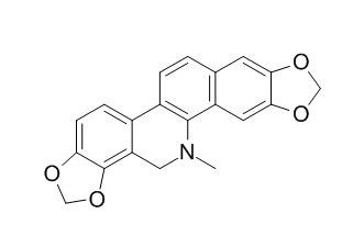
Dihydrosanguinarine
Dihydrosanguinarine has antifungal and anticancer activity.Dihydrosanguinarine at concentrations from 5 microM induced primarily necrosis, whereas apoptosis occurred at 10 microM and above. Dihydrosanguinarine has potential application in the therapy of serious infection caused by I. multifiliis.
Inquire / Order:
manager@chemfaces.com
Technical Inquiries:
service@chemfaces.com
Tel:
+86-27-84237783
Fax:
+86-27-84254680
Address:
1 Building, No. 83, CheCheng Rd., Wuhan Economic and Technological Development Zone, Wuhan, Hubei 430056, PRC
Providing storage is as stated on the product vial and the vial is kept tightly sealed, the product can be stored for up to
24 months(2-8C).
Wherever possible, you should prepare and use solutions on the same day. However, if you need to make up stock solutions in advance, we recommend that you store the solution as aliquots in tightly sealed vials at -20C. Generally, these will be useable for up to two weeks. Before use, and prior to opening the vial we recommend that you allow your product to equilibrate to room temperature for at least 1 hour.
Need more advice on solubility, usage and handling? Please email to: service@chemfaces.com
The packaging of the product may have turned upside down during transportation, resulting in the natural compounds adhering to the neck or cap of the vial. take the vial out of its packaging and gently shake to let the compounds fall to the bottom of the vial. for liquid products, centrifuge at 200-500 RPM to gather the liquid at the bottom of the vial. try to avoid loss or contamination during handling.
Int J Mol Sci.2024, 25(16):8846.
Molecules.2022, 27(19):6681.
Genes (Basel).2021, 12(7):1024.
Int J Mol Sci.2022, 23(21):13406.
J Phys Chem Lett.2021, 12(7):1793-1802.
Hum Exp Toxicol.2023, 42:9603271231171642.
J Separation Science & Technology2016, 51:1579-1588
Anticancer Res.2018, 38(4):2127-2135
Plant Biotechnol (Tokyo).2024, 41(3):267-276.
Research Square2020, doi: 10.21203.
Related and Featured Products
Toxicol In Vitro. 2009 Jun;23(4):580-8.
Cytotoxic activity of sanguinarine and dihydrosanguinarine in human promyelocytic leukemia HL-60 cells.[Pubmed:
19346183]
The benzo[c]phenanthridine alkaloid sanguinarine has been studied for its antiproliferative activity in many cell types. Almost nothing however, is known about the cytotoxic effects of Dihydrosanguinarine, a metabolite of sanguinarine.
METHODS AND RESULTS:
We compared the cytotoxicity of sanguinarine and Dihydrosanguinarine in human leukemia HL-60 cells. Sanguinarine produced a dose-dependent decline in cell viability with IC(50) (inhibitor concentration required for 50% inhibition of cell viability) of 0.9 microM as determined by MTT assay after 4h exposure. Dihydrosanguinarine showed much less cytotoxicity than sanguinarine: at the highest concentration tested (20 microM) and 24h exposure, Dihydrosanguinarine decreased viability only to 52%. Cytotoxic effects of both alkaloids were accompanied by activation of the intrinsic apoptotic pathway since we observed the dissipation of mitochondrial membrane potential, induction of caspase-9 and -3 activities, the appearance of sub-G(1) DNA and loss of plasma membrane asymmetry. This aside, sanguinarine also increased the activity of caspase-8. As shown by flow cytometry using annexin V/propidium iodide staining, 0.5 microM sanguinarine induced apoptosis while 1-4 microM sanguinarine caused necrotic cell death. In contrast, Dihydrosanguinarine at concentrations from 5 microM induced primarily necrosis, whereas apoptosis occurred at 10 microM and above.
CONCLUSIONS:
We conclude that both alkaloids may cause, depending on the alkaloid concentration, both necrosis and apoptosis of HL-60 cells.
Vet Parasitol. 2011 Dec 29;183(1-2):8-13.
Antiparasitic efficacy of dihydrosanguinarine and dihydrochelerythrine from Macleaya microcarpa against Ichthyophthirius multifiliis in richadsin (Squaliobarbus curriculus).[Pubmed:
21813242]
Ichthyophthirius multifiliis is a holotrichous protozoan that invades the gills and skin surfaces of fish and can cause morbidity and high mortality in most species of freshwater fish worldwide. The present study was undertaken to investigate the antiparasitic activity of crude extracts and pure compounds from the leaves of Macleaya microcarpa.
METHODS AND RESULTS:
The chloroform extract showed a promising antiparasitic activity against I. multifiliis. Based on these finding, the chloroform extract was fractionated on silica gel column chromatography in a bioactivity-guided isolation affording two compounds showing potent activity. The structures of the two compounds were elucidated as Dihydrosanguinarine and dihydrochelerythrine by hydrogen and carbon-13 nuclear magnetic resonance spectrum and electron ionization mass spectrometry. The in vivo tests revealed that Dihydrosanguinarine and dihydrochelerythrine were effective against I. multifiliis with median effective concentration (EC(50)) values of 5.18 and 9.43 mg/l, respectively. The acute toxicities (LC(50)) of Dihydrosanguinarine and dihydrochelerythrine for richadsin were 13.3 and 18.2mg/l, respectively.
CONCLUSIONS:
The overall results provided important information for the potential application of Dihydrosanguinarine and dihydrochelerythrine in the therapy of serious infection caused by I. multifiliis.
Food Chem Toxicol. 2008 Jul;46(7):2546-53.
The toxicity and pharmacokinetics of dihydrosanguinarine in rat: a pilot study.[Pubmed:
18495316]
The quaternary benzo[c]phenanthridine alkaloid sanguinarine (SG) is the main component of Sangrovit, a natural livestock feed additive. Dihydrosanguinarine (DHSG) has recently been identified as a SG metabolite in rat. The conversion of SG to DHSG is a likely elimination pathway of SG in mammals.
METHODS AND RESULTS:
This study was conducted to evaluate the toxicity of DHSG in male Wistar rats at concentrations of 100 and 500 ppm DHSG in feed for 90 days (average doses of 14 and 58 mg DHSG/kg body weight/day). No significant alterations in body or organ weights, macroscopic details of organs, histopathology of liver, ileum, kidneys, tongue, heart or gingiva, clinical chemistry or hematology markers in blood in the DHSG-treated animals were found compared to controls. No lymphocyte DNA damage by Comet assay, formation of DNA adducts in liver by 32P-postlabeling, modulation of cytochrome P450 1A1/2 or changes in oxidative stress parameters were found. Thus, repeated dosing of DHSG for 90 days at up to 500 ppm in the diet (i.e. approximately 58 mg/kg/day) showed no evidence of toxicity in contrast to results published in the literature. In parallel, DHSG pharmacokinetics was studied in rat after oral doses 9.1 or 91 mg/kg body weight.
CONCLUSIONS:
The results showed that DHSG undergoes enterohepatic cycling with maximum concentration in plasma at the first or second hour following application. DHSG is cleared from the body relatively quickly (its plasma levels drop to zero after 12 or 18 h, respectively).


