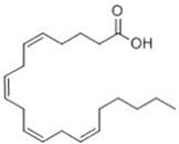
Arachidonic acid
Arachidonic acid is 1 of only 2 unsaturated fatty acids retained in the ovaries of crustaceans and an inhibitor of HR97g, a nuclear receptor expressed in adult ovaries. Arachidonic acid induces retinal arteriolar vasodilation by inhibiting subcellular Ca(2+)-signaling activity in retinal arteriolar myocytes, most likely through a mechanism involving the inhibition of L-type Ca(2+)-channel activity. Arachidonic acid causes an increase in free cytoplasmic calcium concentration ([Ca2+]i) in differentiated skeletal multinucleated myotubes C2C12 and does not induce calcium response in C2C12 myoblasts.
Inquire / Order:
manager@chemfaces.com
Technical Inquiries:
service@chemfaces.com
Tel:
+86-27-84237783
Fax:
+86-27-84254680
Address:
1 Building, No. 83, CheCheng Rd., Wuhan Economic and Technological Development Zone, Wuhan, Hubei 430056, PRC
Providing storage is as stated on the product vial and the vial is kept tightly sealed, the product can be stored for up to
24 months(2-8C).
Wherever possible, you should prepare and use solutions on the same day. However, if you need to make up stock solutions in advance, we recommend that you store the solution as aliquots in tightly sealed vials at -20C. Generally, these will be useable for up to two weeks. Before use, and prior to opening the vial we recommend that you allow your product to equilibrate to room temperature for at least 1 hour.
Need more advice on solubility, usage and handling? Please email to: service@chemfaces.com
The packaging of the product may have turned upside down during transportation, resulting in the natural compounds adhering to the neck or cap of the vial. take the vial out of its packaging and gently shake to let the compounds fall to the bottom of the vial. for liquid products, centrifuge at 200-500 RPM to gather the liquid at the bottom of the vial. try to avoid loss or contamination during handling.
Int J Vet Sci Med.2024, 12(1):134-147.
Current Pharmaceutical Analysis2017, 13(5)
Phytother Res.2020, 34(4):788-795.
Biomedicine & Pharmacotherapy2020, 125:109950
Biochem Biophys Res Commun.2020, 527(4):889-895.
University of East Anglia2023, 93969.
Current Analytical Chemistry2024, 20(8):599-610.
Chinese J of Tissue Engineering Res.2022, 26(17): 2636-2641.
Am J Chin Med.2023, 51(4):1019-1039.
Cancer Sci.2022, 113(4):1406-1416.
Related and Featured Products
Biochemistry (Mosc). 2014 May;79(5):435-9.
Arachidonic acid activates release of calcium ions from reticulum via ryanodine receptor channels in C2C12 skeletal myotubes.[Pubmed:
24954594]
METHODS AND RESULTS:
Arachidonic acid causes an increase in free cytoplasmic calcium concentration ([Ca2+]i) in differentiated skeletal multinucleated myotubes C2C12 and does not induce calcium response in C2C12 myoblasts. The same reaction of myotubes to Arachidonic acid is observed in Ca2+-free medium. This indicates that Arachidonic acid induces release of calcium ions from intracellular stores. The blocker of ryanodine receptor channels of sarcoplasmic reticulum dantrolene (20 µM) inhibits this effect by 68.7 ± 6.3% (p < 0.001). The inhibitor of two-pore calcium channels of endolysosomal vesicles trans-NED19 (10 µM) decreases the response to Arachidonic acid by 35.8 ± 5.4% (p < 0.05). The phospholipase C inhibitor U73122 (10 µM) has no effect.
CONCLUSIONS:
These data indicate the involvement of ryanodine receptor calcium channels of sarcoplasmic reticulum in [Ca2+]i elevation in skeletal myotubes caused by Arachidonic acid and possible participation of two-pore calcium channels from endolysosomal vesicles in this process.
Environ Toxicol Chem. 2015 Mar;34(3):527-35.
Arachidonic acid enhances reproduction in Daphnia magna and mitigates changes in sex ratios induced by pyriproxyfen.[Pubmed:
25393616]
Arachidonic acid is 1 of only 2 unsaturated fatty acids retained in the ovaries of crustaceans and an inhibitor of HR97g, a nuclear receptor expressed in adult ovaries. The authors hypothesized that, as a key fatty acid, Arachidonic acid may be associated with reproduction and potentially environmental sex determination in Daphnia.
METHODS AND RESULTS:
Reproduction assays with Arachidonic acid indicate that it alters female:male sex ratios by increasing female production. This reproductive effect only occurred during a restricted Pseudokirchneriella subcapitata diet. Next, the authors tested whether enriching a poorer algal diet (Chlorella vulgaris) with Arachidonic acid enhances overall reproduction and sex ratios. Arachidonic acid enrichment of a C. vulgaris diet also enhances fecundity at 1.0 µM and 4.0 µM by 30% to 40% in the presence and absence of pyriproxyfen. This indicates that Arachidonic acid is crucial in reproduction regardless of environmental sex determination. Furthermore, the data indicate that P. subcapitata may provide a threshold concentration of Arachidonic acid needed for reproduction. Diet-switch experiments from P. subcapitata to C. vulgaris mitigate some, but not all, of Arachidonic acid's effects when compared with a C. vulgaris-only diet, suggesting that some Arachidonic acid provided by P. subcapitata is retained.
CONCLUSIONS:
In summary, Arachidonic acid supplementation increases reproduction and represses pyriproxyfen-induced environmental sex determination in D. magna in restricted diets. A diet rich in Arachidonic acid may provide protection from some reproductive toxicants such as the juvenile hormone agonist pyriproxyfen.
PLoS One. 2014 Feb 19;9(2):e89347.
Arachidonic acid enhances turnover of the dermal skeleton: studies on zebrafish scales.[Pubmed:
24586706]
In fish nutrition, the ratio between omega-3 and omega-6 poly-unsaturated fatty acids influences skeletal development. Supplementation of fish oils with vegetable oils increases the content of omega-6 fatty acids, such as Arachidonic acid in the diet. Arachidonic acid is metabolized by cyclooxygenases to prostaglandin E2, an eicosanoid with effects on bone formation and remodeling.
METHODS AND RESULTS:
To elucidate effects of poly-unsaturated fatty acids on developing and existing skeletal tissues, zebrafish (Danio rerio) were fed (micro-) diets low and high in Arachidonic acid content. Elasmoid scales, dermal skeletal plates, are ideal to study skeletal metabolism in zebrafish and were exploited in the present study. The fatty acid profile resulting from a high Arachidonic acid diet induced mild but significant increase in matrix resorption in ontogenetic scales of adult zebrafish. Arachidonic acid affected scale regeneration (following removal of ontogenetic scales): mineral deposition was altered and both gene expression and enzymatic matrix metalloproteinase activity changed towards enhanced osteoclastic activity. Arachidonic acid also clearly stimulates matrix metalloproteinase activity in vitro, which implies that resorptive effects of Arachidonic acid are mediated by matrix metalloproteinases.
CONCLUSIONS:
The gene expression profile further suggests that Arachidonic acid increases maturation rate of the regenerating scale; in other words, enhances turnover. The zebrafish scale is an excellent model to study how and which fatty acids affect skeletal formation.
Invest Ophthalmol Vis Sci. 2014 May 2;55(5):2893-902.
Role of ion channels and subcellular Ca2+ signaling in arachidonic acid-induced dilation of pressurized retinal arterioles.[Pubmed:
24699382]
To investigate the mechanisms responsible for the dilatation of rat retinal arterioles in response to Arachidonic acid (AA).
METHODS AND RESULTS:
Changes in the diameter of isolated, pressurized rat retinal arterioles were measured in the presence of AA alone and following pre-incubation with pharmacologic agents inhibiting Ca(2+) sparks and oscillations and K(+) channels. Subcellular Ca(2+) signals were recorded in arteriolar myocytes using Fluo-4-based confocal imaging. The effects of AA on membrane currents of retinal arteriolar myocytes were studied using whole-cell perforated patch clamp recording.
Arachidonic acid dilated pressurized retinal arterioles under conditions of myogenic tone. Eicosatetraynoic acid (ETYA) exerted a similar effect, but unlike AA, its effects were rapidly reversible. Arachidonic acid-induced dilation was associated with an inhibition of subcellular Ca(2+) signals. Interventions known to block Ca(2+) sparks and oscillations in retinal arterioles caused dilatation and inhibited AA-induced vasodilator responses. Arachidonic acid accelerated the rate of inactivation of the A-type Kv current and the voltage dependence of inactivation was shifted to more negative membrane potentials. It also enhanced voltage-activated and spontaneous large-conductance calcium-activated K(+) (BK) currents, but only at positive membrane potentials. Pharmacologic inhibition of A-type Kv and BK currents failed to block AA-induced vasodilator responses. Arachidonic acid suppressed L-type Ca(2+) currents.
CONCLUSIONS:
These results suggest that AA induces retinal arteriolar vasodilation by inhibiting subcellular Ca(2+)-signaling activity in retinal arteriolar myocytes, most likely through a mechanism involving the inhibition of L-type Ca(2+)-channel activity. Arachidonic acid actions on K(+) currents are inconsistent with a model in which K(+) channels contribute to the vasodilator effects of AA.
Apoptosis. 2014 Jul;19(7):1113-27.
Lymphocyte-derived microparticles induce apoptosis of airway epithelial cells through activation of p38 MAPK and production of arachidonic acid.[Pubmed:
24777398 ]
The airway epithelium is critical for the normal integrity and function of the respiratory system. Excessive epithelial cell apoptosis contributes to cell damage and airway inflammation. We previously demonstrated that lymphocyte-derived microparticles (LMPs) induce apoptosis of human bronchial epithelial cells. However, the underlying mechanisms contributing to LMPs-evoked epithelial cell death are largely unknown.
METHODS AND RESULTS:
Here we used bronchial and lung tissue cultures to confirm the pro-apoptotic effects of LMPs. In cell culture experiments, we found that LMPs induced human airway epithelial cell apoptosis with associated increases in caspase-3 activity. In addition, LMPs treatment triggered oxidative stress in epithelial cells by enhancing production of malondialdehyde, superoxide, and reactive oxygen species (ROS), and by inhibiting production of the antioxidant glutathione. Moreover, decreasing cellular ROS with the antioxidant N-acetylcysteine rescued epithelial cell viability. Together, these results demonstrate an important role for oxidative stress in LMPs-induced cell death. In epithelial cells, LMPs treatment induced phosphorylation of p38 MAPK and Arachidonic acid accumulation. Moreover, Arachidonic acid was significantly cytotoxic towards LMPs-treated epithelial cells, whereas inhibition of p38 MAPK was protective against these cytotoxic effects. Similarly, inhibition of Arachidonic acid production led to decreased caspase-3 activity, thus rescuing airway epithelial cells from LMPs-induced cell death.
CONCLUSIONS:
In conclusion, our results show that LMPs induce airway epithelial cell apoptosis by activating p38 MAPK signaling and stimulating production of Arachidonic acid, with consequent increases in oxidative stress and caspase-3 activity. As such, LMPs may be regarded as deleterious markers of epithelial cell damage in respiratory diseases.


