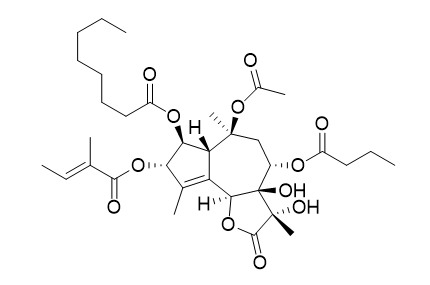
Thapsigargin
Thapsigargin is a potent, non-competitive inhibitor of the sarco/endoplasmic reticulum Ca2+ ATPase (SERCA) with IC50 of 0.353 nM or 0.448 nM for the carbachol-evoked [Ca2+]i-transients with or without a KCl-prestimulation. Thapsigargin induces cell apoptosis. Thapsigargin efficiently inhibits coronavirus (HCoV-229E, MERS-CoV, SARS-CoV-2) replication in different cell types.
Inquire / Order:
manager@chemfaces.com
Technical Inquiries:
service@chemfaces.com
Tel:
+86-27-84237783
Fax:
+86-27-84254680
Address:
1 Building, No. 83, CheCheng Rd., Wuhan Economic and Technological Development Zone, Wuhan, Hubei 430056, PRC
Providing storage is as stated on the product vial and the vial is kept tightly sealed, the product can be stored for up to
24 months(2-8C).
Wherever possible, you should prepare and use solutions on the same day. However, if you need to make up stock solutions in advance, we recommend that you store the solution as aliquots in tightly sealed vials at -20C. Generally, these will be useable for up to two weeks. Before use, and prior to opening the vial we recommend that you allow your product to equilibrate to room temperature for at least 1 hour.
Need more advice on solubility, usage and handling? Please email to: service@chemfaces.com
The packaging of the product may have turned upside down during transportation, resulting in the natural compounds adhering to the neck or cap of the vial. take the vial out of its packaging and gently shake to let the compounds fall to the bottom of the vial. for liquid products, centrifuge at 200-500 RPM to gather the liquid at the bottom of the vial. try to avoid loss or contamination during handling.
Vietnam J. Chemistry2022, 60(2):211-222
Bull. Pharm. Sci., Assiut University2020, 43(2):149-155.
Molecules.2024, 29(21):5161.
Molecules.2021, 26(9):2791.
Food Chem.2021, 360:130063.
Oxid Med Cell Longev.2021, 2021:4883398.
Antioxidants (Basel).2019, 8(8):E307
Plos One.2019, 15(2):e0220084
BMC Plant Biol.2023, 23(1):239.
J Health Sci Med Res.2023, 31584.
Related and Featured Products
Shock . 2017 Apr;47(4):506-513.
Modeling Acute ER Stress in Vivo and in Vitro[Pubmed:
27755507]
The endoplasmic reticulum (ER) is a critical organelle that synthesizes secretory proteins and serves as the main calcium storage site of the cell. The accumulation of unfolded proteins at the ER results in ER stress. Although the association between ER stress and the pathogenesis of many metabolic conditions have been well characterized using both in vivo and in vitro models, no standardized model concerning ER stress exists. Here, we report a standardized model of ER stress using two well-characterized ER stress-inducing agents, Thapsigargin and tunicamycin. Our aim in this current study was 2-fold: to characterize and establish which agent is optimal for in vitro use to model acute ER stress and to evaluate which agent is optimal for in vivo use. To study the first aim we used two well-established metabolic cell lines; human hepatocellular carcinoma (HepG2s) and differentiated mouse adipocytes (3T3-L1). In the second aim we utilized C57BL/6J mice that were randomized into three treatment groups of sham, Thapsigargin, and tunicamycin. Our in vitro results showed that tunicamycin worked as a rapid and efficacious inducer of ER stress in adipocytes consistently, whereas Thapsigargin and tunicamycin were equally effective in inducing ER stress in hepatocytes. In regards to our in vivo results, we saw that tunicamycin was superior in not only inducing ER stress but also recapturing the metabolic alterations associated with ER stress. Thus, our findings will help guide and inform researchers as to which ER stress agent is appropriate with regards to their model.
Brain Res . 2004 Jun 18;1011(2):177-186
Differential thapsigargin-sensitivities and interaction of Ca2+ stores in human SH-SY5Y neuroblastoma cells[Pubmed:
15157804]
In human SH-SY5Y neuroblastoma cells, two distinct intracellular Ca2+ stores, a KCl-/caffeine-sensitive and a carbachol-/IP3-sensitive store, were demonstrated previously. In this study, responses of these two intracellular Ca2+ stores to Thapsigargin were characterized. Ca2+-release from these stores was evoked either by high K+ (100 mM KCl) or by 1 mM carbachol, and changes in the intracellular Ca2+ level were monitored using Fura-2 fluorimetry. A sequential stimulation protocol (KCl-->carbachol or vice versa) allowed evaluation of the individual contribution of different Ca2+ stores to the evoked intracellular Ca2+ ([Ca2+]i)-transients and the dynamic interaction between them. Thapsigargin (0.05 nM - 20 microM) alone induced a [Ca2+]i-transient. Both the carbachol- and the KCl-evoked [Ca2+]i-transients were inhibited by Thapsigargin, but with very different sensitivities. Thapsigargin inhibited the carbachol-evoked [Ca2+]i-transients with (IC50 = 0.353 nM) or without (IC50 = 0.448 nM) a KCl-prestimulation, but an additional small component, with a much lower sensitivity (IC50=4814 nM), was observed in the absence of a KCl-prestimulation. In contrast, the KCl-evoked [Ca2+]i-transients displayed only one component with a very low sensitivity to Thapsigargin in both absence (IC50=3343 nM) and presence (IC50=6858 nM) of a carbachol-prestimulation. These findings suggest that the sarco-/endoplasmic reticular Ca2+ ATPases associated with the KCl-/caffeine- and carbachol-/IP3-sensitive intracellular Ca2+ stores differ from each other, either in types or in their post-translational modification. Such difference might play important role in the regulation of neuronal Ca2+ homeostasis.
Biochem J . 1995 Jan 15;305 ( Pt 2)(Pt 2):525-528.
Thapsigargin inhibits Ca2+ entry into human neutrophil granulocytes[Pubmed:
7832770]
The mechanism of Ca2+ entry after ligand binding to receptors on the surface of non-excitable cells is a current focus of interest. Considerable attention has been given to Ca2+ influx induced by emptying of intracellular pools. Thapsigargin, an inhibitor of microsomal Ca(2+)-ATPase, is an important tool in inducing store-regulated Ca2+ influx. In the present paper we show that, at concentrations above 500 nM, Thapsigargin also has an opposite effect: it inhibits store-regulated Ca2+ influx into Fura-2-loaded human neutrophil granulocytes. As Thapsigargin has been frequently applied at concentrations up to 2 microM, its inhibitory action on plasma-membrane Ca2+ fluxes deserves consideration.
Shock . 2017 Apr;47(4):506-513.
Modeling Acute ER Stress in Vivo and in Vitro[Pubmed:
27755507]
The endoplasmic reticulum (ER) is a critical organelle that synthesizes secretory proteins and serves as the main calcium storage site of the cell. The accumulation of unfolded proteins at the ER results in ER stress. Although the association between ER stress and the pathogenesis of many metabolic conditions have been well characterized using both in vivo and in vitro models, no standardized model concerning ER stress exists. Here, we report a standardized model of ER stress using two well-characterized ER stress-inducing agents, Thapsigargin and tunicamycin. Our aim in this current study was 2-fold: to characterize and establish which agent is optimal for in vitro use to model acute ER stress and to evaluate which agent is optimal for in vivo use. To study the first aim we used two well-established metabolic cell lines; human hepatocellular carcinoma (HepG2s) and differentiated mouse adipocytes (3T3-L1). In the second aim we utilized C57BL/6J mice that were randomized into three treatment groups of sham, Thapsigargin, and tunicamycin. Our in vitro results showed that tunicamycin worked as a rapid and efficacious inducer of ER stress in adipocytes consistently, whereas Thapsigargin and tunicamycin were equally effective in inducing ER stress in hepatocytes. In regards to our in vivo results, we saw that tunicamycin was superior in not only inducing ER stress but also recapturing the metabolic alterations associated with ER stress. Thus, our findings will help guide and inform researchers as to which ER stress agent is appropriate with regards to their model.
bioRxiv 2020.08.26.266304.
Inhibiting coronavirus replication in cultured cells by chemical ER stress[Reference:
WebLink]
Coronaviruses (CoVs) are important human pathogens for which no specific treatment is available. Here, we provide evidence that pharmacological reprogramming of ER stress pathways can be exploited to suppress CoV replication. We found that the ER stress inducer Thapsigargin efficiently inhibits coronavirus (HCoV-229E, MERS-CoV, SARS-CoV-2) replication in different cell types, (partially) restores the virus-induced translational shut-down, and counteracts the CoV-mediated downregulation of IRE1α and the ER chaperone BiP. Proteome-wide data sets revealed specific pathways, protein networks and components that likely mediate the Thapsigargin-induced antiviral state, including HERPUD1, an essential factor of ER quality control, and ER-associated protein degradation complexes. The data show that Thapsigargin hits a central mechanism required for CoV replication, suggesting that Thapsigargin (or derivatives thereof) may be developed into broad-spectrum anti-CoV drugs.
One Sentence Summary / Running title Suppression of coronavirus replication through Thapsigargin-regulated ER stress, ERQC / ERAD and metabolic pathways
ScientificWorldJournal . 2014 Feb 9;2014:605416.
Effects of thapsigargin on the proliferation and survival of human rheumatoid arthritis synovial cells[Pubmed:
24688409]
A series of experiments have been carried out to investigate the effects of different concentrations of Thapsigargin (0, 0.001, 0.1, and 1 μM) on the proliferation and survival of human rheumatoid arthritis synovial cells (MH7A). The results showed that Thapsigargin can block the cell proliferation in human rheumatoid arthritis synovial cells in a time- and dose-dependent manner. Results of Hoechst staining suggested that Thapsigargin may induce cell apoptosis in MH7A cells in a time- and dose-dependent manner, and the percentages of cell death reached 44.6% at Thapsigargin concentration of 1 μM treated for 4 days compared to the control. The protein and mRNA levels of cyclin D1 decreased gradually with the increasing of Thapsigargin concentration and treatment times. Moreover, the protein levels of mTORC1 downstream indicators pS6K and p4EBP-1 were reduced by Thapsigargin treatment at different concentrations and times, which should be responsible for the reduced cyclin D1 expressions. Our results revealed that Thapsigargin may effectively impair the cell proliferation and survival of MH7A cells. The present findings will help to understand the molecular mechanism of fibroblast-like synoviocytes proliferations and suggest that Thapsigargin is of potential for the clinical treatment of rheumatoid arthritis.


