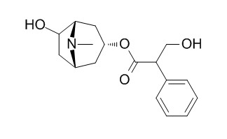
Anisodamine
Anisodamine, an anticholinergic drug, has antishock effect, which is intimately linked to alpha7nAChR-dependent anti-inflammatory pathway. Anisodamine demonstrates a direct cardiac depressive action at the myocyte level, which may be related to, at least in part, NO production and cholinoceptor antagonism, it causes the changes of structure and function in the transmembrane domain of the Ca(2+)-ATPase from sarcoplasmic reticulum. Anisodamine, a vasoactive drug, can abate endogenous endotoxaemia subsequent to splanchnic vasoconstriction due to hypovolaemia, it alleviates inflammatory damage by significantly reducing the expressions of VEGF and ICAM-1, and shows significant protective effects in an animal model of infusion phlebitis. Anisodamine also inhibits shiga toxin type 2-mediated tumor necrosis factor-alpha production in vitro and in vivo.
Inquire / Order:
manager@chemfaces.com
Technical Inquiries:
service@chemfaces.com
Tel:
+86-27-84237783
Fax:
+86-27-84254680
Address:
1 Building, No. 83, CheCheng Rd., Wuhan Economic and Technological Development Zone, Wuhan, Hubei 430056, PRC
Providing storage is as stated on the product vial and the vial is kept tightly sealed, the product can be stored for up to
24 months(2-8C).
Wherever possible, you should prepare and use solutions on the same day. However, if you need to make up stock solutions in advance, we recommend that you store the solution as aliquots in tightly sealed vials at -20C. Generally, these will be useable for up to two weeks. Before use, and prior to opening the vial we recommend that you allow your product to equilibrate to room temperature for at least 1 hour.
Need more advice on solubility, usage and handling? Please email to: service@chemfaces.com
The packaging of the product may have turned upside down during transportation, resulting in the natural compounds adhering to the neck or cap of the vial. take the vial out of its packaging and gently shake to let the compounds fall to the bottom of the vial. for liquid products, centrifuge at 200-500 RPM to gather the liquid at the bottom of the vial. try to avoid loss or contamination during handling.
Exp Neurobiol.2018, 27(3):200-209
Phytomedicine.2019, 65:153089
Curr Res Food Sci.2024, 9:100827.
J Health Sci Med Res.2023, 31584.
J Food Biochem.2021, 45(7):e13774.
Oxid Med Cell Longev2019, 9056845:13
J Appl Biol Chem.2024, 67:39,281-288.
Phytomedicine.2018, 41:62-66
Biomolecules.2024, 14(4):451.
Evid Based Complement Alternat Med.2019, 2019:2135351
Related and Featured Products
Burns. 1997 Mar;23(2):142-6.
Anisodamine restores bowel circulation in burn shock.[Pubmed:
9177881]
METHODS AND RESULTS:
In a group of eight burn patients with a mean of 65.3 +/- 17.4 per cent TBSA burn injury (range 50-90 per cent TBSA), accompanied by a mean of 43.5 +/- 18.9 per cent TBSA full-thickness injury, it was shown that the evidence of global hypovolaemia had disappeared at 12 h after the injury following aggressive fluid resuscitation, while there was still a subnormal pHi of stomach at 48 h. As a prolonged period of inadequacy of oxygen delivery to the intestine might result in impairment of the intestinal mucosal barrier function, and then endogenous endotoxaemia might ensue, it seems to be important to correct intestinal hypoxia as early as possible. Since the inadequate perfusion to the gut wall is due to selective vasoconstriction of the mesenteric vasculature, logic dictates that the use of a vasodilator is in order. Anisodamine, an anticholinergic drug, was then given in six burn patients with comparable burn size and amount of fluid replenishment with the eight patients in the control group. It was clearly demonstrated that gastric pHi returned to normal before 48 h after injury. Plasma endotoxin and TNF contents were measured, and they were significantly lower than control values after 72 h.
CONCLUSIONS:
In conclusion, it is believed that Anisodamine might be a valuable adjunct to the resuscitation regime of burn shock, and, therefore, a promising drug to abate endogenous endotoxaemia subsequent to splanchnic vasoconstriction due to hypovolaemia. The shortcomings of the drug were a mild abdominal distention and tachycardia after its administration.
Exp Biol Med (Maywood). 2001 Jun;226(6):597-604.
Anisodamine inhibits shiga toxin type 2-mediated tumor necrosis factor-alpha production in vitro and in vivo.[Pubmed:
11395932]
Cytokines, in particular tumor necrosis factor (TNF), appear to be necessary to develop the pathological process of Shiga toxin-producing Escherichia coli (STEC) infection.
METHODS AND RESULTS:
In this study we examined the effect of Anisodamine, a vasoactive drug, on TNF-alpha production in Shiga toxin type 2 (Stx2)-stimulated human monocytic cells in vitro and in Stx2-injected mice sera in vivo. Human monocytes and THP-1 cells were stimulated by Stx2 (1-100 ng/ml) with or without Anisodamine addition (1-400 micrograms/ml). For in vivo evaluations, C57BL/6 mice were given a single intraperitoneal injection of Anisodamine (6-50 mg/kg) or saline after intraperitoneal injection of Stx2 (50 ng/kg). The results showed that Anisodamine suppressed Stx2-induced TNF-alpha production in a dose- and time-dependent manner. Anisodamine also suppressed Stx2-induced TNF-alpha mRNA expression. Further study showed that endogenous prostaglandin E2 may be involved in this inhibitory effect. In contrast to TNF-alpha mRNA, Anisodamine at concentrations as high as 400 micrograms/ml did not decrease Stx2-induced IL-1 beta and IL-8 mRNA levels. In addition, Anisodamine (> 50 micrograms/ml) increased Stx2-stimulated THP-1 cell viability. Levels of TNF-alpha in Anisodamine-treated mice sera were significantly lower than those in the saline-treated group 1.5 and 24 hr after Stx2 injection. Anisodamine induced a lower percentage of death in Stx2-injected mice.
CONCLUSIONS:
Taken together, our results indicate that Anisodamine has an important regulatory effect on Stx2-induced TNF-alpha production in vitro and in vivo. The present study suggested that this drug should be further investigated for its effects on Stx2-mediated diseases in humans.
Chin Med J (Engl). 2012 Jan;125(2):300-5.
Effects of anisodamine on the expressions of vascular endothelial growth factor and intercellular adhesion molecule 1 in experimental infusion phlebitis.[Pubmed:
22340563]
Infusion phlebitis is the most common side effect of clinical intravenous drug therapy and several clinical studies have demonstrated that Anisodamine can effectively prevent the occurrence of infusion phlebitis. This study was designed to investigate effects of Anisodamine on the expressions of vascular endothelial growth factor (VEGF) and intercellular adhesion molecule 1 (ICAM-1) in a rabbit model of infusion phlebitis and to analyze the mechanisms of Anisodamine effect on the prevention and treatment of experimental infusion phlebitis.
METHODS AND RESULTS:
Twenty-four specific pathogen-free male Japanese white rabbits were randomly assigned to the control group, the model group, the magnesium sulfate group and the Anisodamine group. The rabbit model of infusion phlebitis, induced by intravenous administration, was established and expressions of VEGF and ICAM-1 were determined and contrasted with the control group treated with normal saline. We evaluated expression by histopathology, immunohistochemistry, reverse transcription-polymerase chain reaction, and Western blotting assay.Pathohistological changes of the model group were observed, such as loss of venous endothelial cells, inflammatory cell infiltration, edema and thrombus. The magnesium sulfate group and the Anisodamine group showed significant protective effects on vascular congestion, inflammatory cell infiltration, proliferation, swelling of endothelium and perivascular hemorrhage. The model group showed the highest expressions of VEGF and ICAM-1 of the four groups (P < 0.01). On the contrary, Anisodamine alleviated the inflammatory damage by significantly reducing the expressions of VEGF and ICAM-1 compared with the model group (P < 0.01). There was no significant difference in the expressions of VEGF and ICAM-1 between the magnesium sulfate group and the Anisodamine group (P > 0.05).
CONCLUSIONS:
Anisodamine alleviates inflammatory damage by significantly reducing the expressions of VEGF and ICAM-1, and shows significant protective effects in an animal model of infusion phlebitis.
Biosci Biotechnol Biochem. 2004 Jan;68(1):126-31.
Anisodamine causes the changes of structure and function in the transmembrane domain of the Ca(2+)-ATPase from sarcoplasmic reticulum.[Pubmed:
14745174]
CONCLUSIONS:
The effects of Anisodamine on the Ca(2+)-ATPsae of sarcoplasmic reticulum (SR) were investigated by using differential scanning calorimetry to measure the ability of Anisodamine to denature the transmembrane domain and the cytoplasmic domain. Anisodamine significantly altered the thermotropic phase behaviors of the transmembrane domain of purified Ca(2+)-ATPase. Specifically, the melting temperature of the transmembrane domain moved toward lower temperatures with the concentrations of Anisodamine increasing and the thermotropic phase peak was abolished at 10 mM, indicating that the stabilized structure of the transmembrane domain in the presence of Ca2+ could be destabilized by Anisodamine. Decreases of the intrinsic fluorescence and increases of the extrinsic fluorescence of ANS, a fluorescent probe, showed the exposure of tryptophan and hydrophobic region, respectively, suggesting again that Anisodamine caused a less compact conformation in the transmembrane domain. A marked inhibition of the Ca2+ uptake activity of SR Ca(2+)-ATPase was observed when the addition of Anisodamine. The drug did not affect the cytoplasmic domain of the enzyme and only slightly decreased the ATPase activity of the enzyme at concentrations up to 10 mM. This was likely due to the destabilized protein transmembrane domain.
CONCLUSIONS:
To sum up, our results revealed that Anisodamine interacted specifically with the transmembrane domain of SR Ca(2+)-ATPase and inhibited the Ca2+ uptake activity of the enzyme.
Crit Care Med. 2009 Feb;37(2):634-41.
Antishock effect of anisodamine involves a novel pathway for activating alpha7 nicotinic acetylcholine receptor.[Pubmed:
19114896 ]
METHODS AND RESULTS:
Sprague-Dawley rats were injected with lipopolysaccharide (LPS) (15 mg/kg, intravenous) to induce septic shock.
Methyllycaconitine, a selective alpha7nAChR antagonist, was administered (10 mg/kg, intraperitoneal) 10 minutes before Anisodamine (10 mg/kg, intravenous). Mean arterial pressure was monitored and cytokines were analyzed 2 hours after the onset of LPS. In vagotomized mice and alpha7nAChR-deficient mice, the antishock effect of Anisodamine was appraised, respectively. RAW264.7 cells were stained by fluorescein isothiocyanate- labeled-alpha-bungarotoxin and the fluorescence intensity was observed. Mice peritoneal macrophages were pretreated and stimulated with LPS, and tumor necrosis factor (TNF)-alpha in the supernatant was measured by enzyme-linked immunosorbent assay. Methyllycaconitine significantly antagonized the beneficial effect of Anisodamine on mean arterial pressure and TNF-alpha, interleukin-1beta expression in response to LPS. The antishock effects of Anisodamine were markedly attenuated in vagotomized mice and alpha7nAChR-deficient mice. In vitro, Anisodamine significantly augmented the effect of acetylcholine on fluorescence intensity stained with fluorescein isothiocyanate-labeled-alpha-bungarotoxin and TNF-alpha production stimulated with LPS.
CONCLUSIONS:
These findings demonstrate that the antishock effect of Anisodamine is intimately linked to alpha7nAChR-dependent anti-inflammatory pathway.
Eur J Pharmacol. 2002 Mar 29;439(1-3):21-5.
Anisodamine inhibits cardiac contraction and intracellular Ca(2+) transients in isolated adult rat ventricular myocytes.[Pubmed:
11937088]
Increased cardiac workload often leads to serious complications during cardiac surgery such as pericardiopulmonary bypass. Various agents have been applied to lower peripheral resistance and cardiac workload, one of which, Anisodamine, is widely used in Asia. However, the direct action of Anisodamine on cardiac contractile property is essentially unknown.
METHODS AND RESULTS:
This study was designed to examine the influence of Anisodamine on ventricular contractile function at the single cardiac myocyte level. Ventricular myocytes from adult rat hearts were stimulated to contract at 0.5 Hz, and mechanical and intracellular Ca(2+) properties were evaluated using an IonOptix Myocam system. Contractile properties analyzed included peak shortening (PS), time-to-PS (TPS), time-to-90% relengthening (TR(90)), maximal velocity of shortening/relengthening (+/-dL/dt), intracellular Ca(2+) fluorescence intensity change (DeltaFFI) and decay (tau). Anisodamine exhibited a concentration-dependent (10(-12)-10(-6) M) inhibition in PS and DeltaFFI, with maximal inhibitions of 44.7% and 47.2%, respectively. Anisodamine inhibited +/-dL/dt, lowered resting FFI but elicited no effect on TPS/TR(90) and tau. Pretreatment with the nitric oxide synthase (NOS) inhibitor N(omega)-nitro-L-arginine methyl ester (L-NAME, 100 microM) abolished the inhibitory effect of Anisodamine in cell shortening. In addition, Anisodamine prevented cholinoceptor agonist carbachol-induced positive cardiac contractile response.
CONCLUSIONS:
This study demonstrated a direct cardiac depressive action of Anisodamine at the myocyte level, which may be related to, at least in part, NO production and cholinoceptor antagonism.
Am J Chin Med. 2011;39(5):853-66.
Inhibition of endoplasm reticulum stress by anisodamine protects against myocardial injury after cardiac arrest and resuscitation in rats.[Pubmed:
21905277 ]
Anisodamine is a multi-functional bio-alkaloid with vascular activity. Our previous studies have revealed that Anisodamine protects the heart from ischemia/reperfusion (I/R) injury induced by cardiac arrest (CA) and resuscitation. This study aimed to explore whether the protective effect of Anisodamine is mediated by inhibition of the endoplasmic reticulum stress (ERS) response, which has been demonstrated to implicate in various I/R injuries.
METHODS AND RESULTS:
After 5 min of CA induced by electric stimulation, Wistar rats were randomly selected to receive cardiopulmonary resuscitation (CPR, including chest compression and epinephrine infusion) with or without Anisodamine injection (n = 50/group). Hearts were harvested 24 h after the return of spontaneous circulation (ROSC). Sham-operated animals served as non-ischemic controls (n = 10). The survival rate, cardiomyocyte apoptosis, and the protein expression of ERS markers were detected. Thirty-three of the 50 rats in the Ani + CA/R group were successfully resuscitated, whereas only 18 of the 50 rats in the CA/R group gained ROSC. Survival to 24 h was significantly improved in the Anisodamine treatment group (Ani + CA/R, n = 22/50) compared to the group with standard CPR (CA/R, n = 8/50). Anisodamine markedly decreased the number of apoptotic cardiomyocytes, the protein expression of GRP78, CHOP, and the active form of Caspase3 compared to the CA/R group.
CONCLUSIONS:
Our data suggest that Anisodamine protects against cellular damage in rat hearts after CA and resuscitation, at least in part, by inhibiting myocardial ERS.


